
Drug Information
General Information of This Drug
| Drug ID | DRG00405 | |||||
|---|---|---|---|---|---|---|
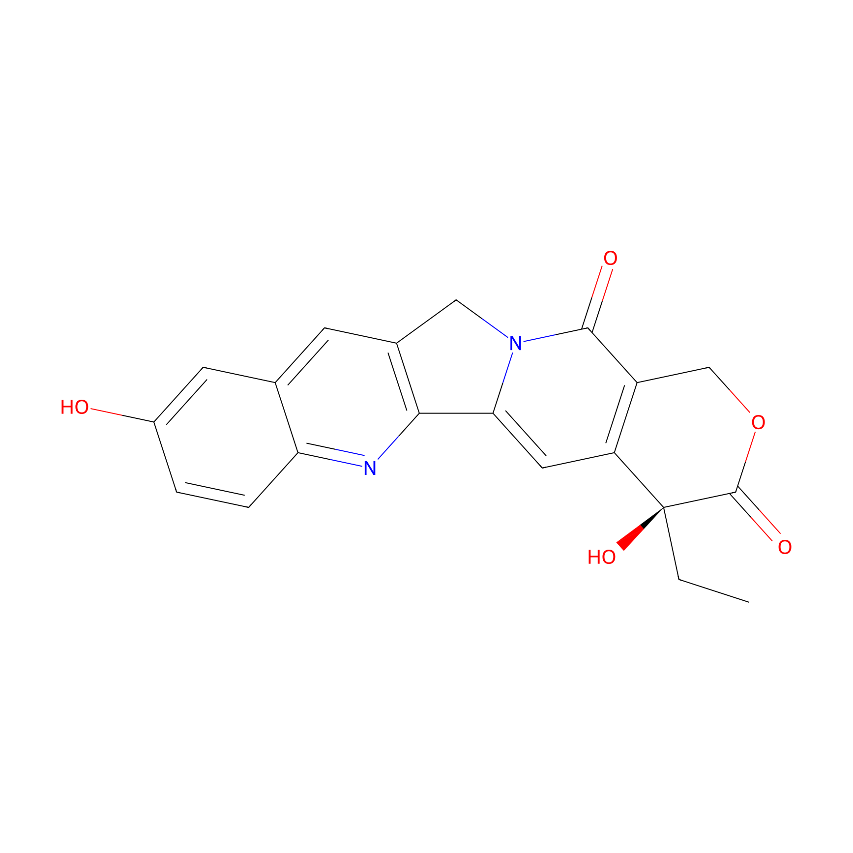
| Drug Name | 10-Hydroxycamptothecin | |||||
| Synonyms |
10-Hydroxycamptothecin; 19685-09-7; (S)-10-Hydroxycamptothecin; Hydroxycamptothecin; 10-hydroxycamptothecine; 10-Hydroxy camptothecin; (S)-4-Ethyl-4,9-dihydroxy-1H-pyrano[3',4':6,7]indolizino[1,2-b]quinoline-3,14(4H,12H)-dione; Hydroxycamptothecine; (4S)-4-Ethyl-4,9-dihydroxy-1H-pyrano[3',4':6,7]indolizino[1,2-b]quinoline-3,14(4H,12H)-dione; Camptothecin, hydroxy-; Camptothecine, 10-hydroxy-; 10-HCPT; 10-Hydroxy-Camptothecin; NSC107124; NSC-107124; CHEMBL273862; CHEBI:81395; 9Z01632KRV; Hydroxy camptothecine; MFCD02093100; (S)-4-ethyl-4,9-dihydroxy-1,12-dihydro-14H-pyrano[3',4':6,7]indolizino[1,2-b]quinoline-3,14(4H)-dione; Camptothecin, 10-hydroxy-; (20S)-4-Ethyl-4,9-dihydroxy-1,12-dihydro-4H-2-oxa-6,12a-diaza-dibenzo[b,h]fluorene-3,13-dione; (S)-4-Ethyl-4,9-dihydroxy-1H-pyrano[3',4':6,7]indolizino[1,2-b]quinoline-3,14(4H,12H)-dione ((S)-10-Hydroxycamptothecin); NSC 107124; (S)-10-Hydroxycamptothecin hydrate; UNII-9Z01632KRV; CAMPTOTHECIN, 10-HYDROXY; 10-Hydroxy-CPT; Spectrum_001639; (+)-(S)-10-HYDROXYCAMPTOTHECIN; SpecPlus_000763; Spectrum2_001660; Spectrum3_001621; Spectrum4_001815; Spectrum5_000549; ethyl(dihydroxy)[?]dione; SCHEMBL25875; BSPBio_003281; KBioGR_002454; KBioSS_002119; 1H-Pyrano(3',4':6,7)indolizino(1,2-b)quinoline-3,14(4H,12H)-dione, 4-ethyl-4,9-dihydroxy-, hydrate, (S)-; DivK1c_006859; SPECTRUM1504123; SPBio_001819; KBio1_001803; KBio2_002119; KBio2_004687; KBio2_007255; KBio3_002501; DTXSID00941444; EX-A988; BCP01385; HY-N0095; (+)-10-HYDROXYCAMPTOTHECIN; BDBM50008922; CCG-38770; s2423; s3898; AKOS015919293; AC-5502; BCP9000058; CS-5193; DB12385; 10-HYDROXYCAMPTOTHECIN [WHO-DD]; NCGC00095986-01; NCGC00095986-02; NCGC00095986-03; NCGC00095986-04; NCGC00178165-01; 1H-Pyrano(3',4':6,7)indolizino(1,2-b)quinoline-3,14(4H,12H)-dione-,4-ethyl-4,9-dihydroxy-, (S)-; AC-13221; AS-13196; NCI60_000173; SY010687; H1463; NS00018317; A25382; C17939; EN300-19810684; SR-05000002620; Q-100241; SR-05000002620-1; BRD-K63784565-001-02-1; BRD-K63784565-001-03-9; BRD-K63784565-001-05-4; BRD-K63784565-001-06-2; Q27155328; Z3093896188; 4-Ethyl-4,9-dihydroxy-1,12-dihydro-4H-2-oxa-6,12a-diaza-dibenzo[b,h]fluorene-3,13-dione; (19S)-19-ethyl-7,19-dihydroxy-17-oxa-3,13-diazapentacyclo[11.8.0.0^{2,11}.0^{4,9}.0^{15,20}]henicosa-1(21),2(11),3,5,7,9,15(20)-heptaene-14,18-dione; (19S)-19-ethyl-7,19-dihydroxy-17-oxa-3,13-diazapentacyclo[11.8.0.02,11.04,9.015,20]henicosa-1(21),2(11),3,5,7,9,15(20)-heptaene-14,18-dione; (S)-10-Hydroxycamptothecin;-;(+/-)-4-ethyl-4,9-dihydroxy-1h-pyrano[3',4':6,7]indolizino[1,2-b]quinoline-3,14(4h,12h)-dione; (S)-4-Ethyl-4,9-dihydroxy-1H-pyrano[3 inverted exclamation mark ,4 inverted exclamation mark :6,7]indolizino[1,2-b]quinoline-3,14-(4H,12H)-dione; (S)-4-Ethyl-4,9-dihydroxy-1H-pyrano[3',4':6,7]indolizino[1,2-b]quinoline-3,14-(4H,12H)-dione; 1H-PYRANO(3',4':6,7)INDOLIZINO(1,2-B)QUINOLINE-3,14(4H,12H)-DIONE, 4-ETHYL-4,9-DIHYDROXY-, (4S)-; 1H-Pyrano[3',7]indolizino[1,2-b]quinoline-3,14(4H,12H)-dione-,4-ethyl-4,9-dihydroxy-, (S)-; 4-Ethyl-4,10-dihydroxy-1,12-dihydro-4H-2-oxa-6,12a-diaza-dibenzo[b,h]fluorene-3,13-dione; 4-Ethyl-4,9-dihydroxy-1,12-dihydro-4H-2-oxa-6,12a-diaza-dibenzo[b,h]fluorene-3,13-dione (10-hydroxycamptothecin)
Click to Show/Hide
|
|||||
| Target(s) | DNA topoisomerase 1 (TOP1) | Target Info | ||||
| Structure |
|
|||||
| Formula |
C20H16N2O5
|
|||||
| #Ro5 Violations (Lipinski): 0 | Molecular Weight (mw) | 364.4 | ||||
| Lipid-water partition coefficient (xlogp) | 0.6 | |||||
| Hydrogen Bond Donor Count (hbonddonor) | 2 | |||||
| Hydrogen Bond Acceptor Count (hbondacc) | 6 | |||||
| Rotatable Bond Count (rotbonds) | 1 | |||||
| PubChem CID | ||||||
| Canonical smiles |
CCC1(C2=C(COC1=O)C(=O)N3CC4=C(C3=C2)N=C5C=CC(=CC5=C4)O)O
|
|||||
| InChI |
InChI=1S/C20H16N2O5/c1-2-20(26)14-7-16-17-11(5-10-6-12(23)3-4-15(10)21-17)8-22(16)18(24)13(14)9-27-19(20)25/h3-7,23,26H,2,8-9H2,1H3/t20-/m0/s1
|
|||||
| InChIKey |
HAWSQZCWOQZXHI-FQEVSTJZSA-N
|
|||||
| IUPAC Name |
(19S)-19-ethyl-7,19-dihydroxy-17-oxa-3,13-diazapentacyclo[11.8.0.02,11.04,9.015,20]henicosa-1(21),2(11),3,5,7,9,15(20)-heptaene-14,18-dione
|
|||||
The activity data of This Drug
| Standard Type | Value | Disease Model | Cell line | Cell line ID | Ref. | |
|---|---|---|---|---|---|---|
| Half Maximal Inhibitory Concentration (IC50) | 18 nM | Lymphoblastic leukemia | L1210 cell | CVCL_0382 | [1] | |
| Half Maximal Inhibitory Concentration (IC50) | 110 nM | Lung adenocarcinoma | A-549 cell | CVCL_0023 | [2] | |
| Half Maximal Inhibitory Concentration (IC50) | 140 nM | Hepatocellular carcinoma | SMMC-7721 cell | CVCL_0534 | [3] | |
| Half Maximal Inhibitory Concentration (IC50) | 148 nM | Colon carcinoma | HCT 116 cell | CVCL_0291 | [4] | |
| Half Maximal Inhibitory Concentration (IC50) | 250 nM | Acute myeloid leukemia | HL-60 cell | CVCL_0002 | [5] | |
| Half Maximal Inhibitory Concentration (IC50) | 300 nM | Lung adenocarcinoma | A-549 cell | CVCL_0023 | [5] | |
| Half Maximal Inhibitory Concentration (IC50) | 600 nM | Human papillomavirus-related endocervical adenocarcinoma | KB cell | CVCL_0372 | [6] | |
| Half Maximal Inhibitory Concentration (IC50) | 700 nM | Ovarian endometrioid adenocarcinoma | A2780 cell | CVCL_0134 | [6] | |
| Half Maximal Inhibitory Concentration (IC50) | 800 nM | Lung adenocarcinoma | A-549 cell | CVCL_0023 | [7] | |
| Half Maximal Inhibitory Concentration (IC50) | 1.75 uM | Lung adenocarcinoma | A-549 cell | CVCL_0023 | [8] | |
| Half Maximal Inhibitory Concentration (IC50) | 2.4 uM | Invasive breast carcinoma | MCF-7 cell | CVCL_0031 | [7] | |
| Half Maximal Inhibitory Concentration (IC50) | 2.47 uM | Hepatoma | Bel-7402 cell | CVCL_5492 | [4] | |
| Half Maximal Inhibitory Concentration (IC50) | 3.2 uM | Colon carcinoma | HCT 116 cell | CVCL_0291 | [9] | |
| Half Maximal Inhibitory Concentration (IC50) | 3.5 uM | Colon carcinoma | HCT 116 cell | CVCL_0291 | [7] | |
| Half Maximal Inhibitory Concentration (IC50) | 6.55 uM | Gastric cancer | MGC-803 cell | CVCL_5334 | [10] | |
| Half Maximal Inhibitory Concentration (IC50) | 10.1 uM | Hepatoblastoma | Hep-G2 cell | CVCL_0027 | [7] | |
| Half Maximal Inhibitory Concentration (IC50) | 10.21 uM | Hepatoblastoma | Hep-G2 cell | CVCL_0027 | [11] | |
| Half Maximal Inhibitory Concentration (IC50) | 11.83 uM | Bladder carcinoma | T24 cell | CVCL_0554 | [8] | |
| Half Maximal Inhibitory Concentration (IC50) | 20.4 uM | Cervical carcinoma | L02 cell | CVCL_6926 | [9] | |
| Half Maximal Inhibitory Concentration (IC50) | 48.2 uM | Invasive breast carcinoma | MCF-7 cell | CVCL_0031 | [12] | |
Each Peptide-drug Conjugate Related to This Drug
Full Information of The Activity Data of The PDC(s) Related to This Drug
R-L-HCPT [Investigative]
Discovered Using Cell Line-derived Xenograft Model
| Experiment 1 Reporting the Activity Data of This PDC | [13] | ||||
| Indication | Solid tumor | ||||
| Efficacy Data | Tumer volume | 700 mm3 | |||
| Administration Time | 10 days | ||||
| MOA of PDC |
Our previous studies demonstrated that R-lycosin-I was a typical cationic anticancer peptide that contained a high relative abundance of positively charged arginine residues and possessed an amphiphilic character to allow its assembly into nanostructures in aqueous environments. Herein, we designed and synthesized a self-assembling anticancer conjugate in which R-lycosin-I covalently coupled with HCPT, a DNA topoisomerase I inhibitor through the glutamic anhydride linker. This conjugate (R-L-HCPT) could spontaneously associate into uniform 40-60 nm nanospheres in aqueous solution. The R-L-HCPT nanospheres exhibited excellent antitumor growth activity and antimetastatic efficacy compared with free HCPT or free R-lycosin-I in vitro and in vivo. Our study might provide new opportunities for the development of a self-assembling peptide-drug delivery system that could synergistically enhance anticancer outcomes.
Click to Show/Hide
|
||||
| Description |
Compared with the tumors in the control group with a final tumor volume of 3800 mm3, the tumors in the R-L-HCPT group grew much more slowly, and the mean tumor volume expanded from 200 to 700 mm3 within 10 days.
|
||||
| In Vivo Model | Murine melanoma B16-F10 xenograft model in BALB/c mice. | ||||
| Experiment 2 Reporting the Activity Data of This PDC | [13] | ||||
| Indication | Solid tumor | ||||
| Efficacy Data | Body weight | 31g | |||
| Administration Time | 10 days | ||||
| MOA of PDC |
Our previous studies demonstrated that R-lycosin-I was a typical cationic anticancer peptide that contained a high relative abundance of positively charged arginine residues and possessed an amphiphilic character to allow its assembly into nanostructures in aqueous environments. Herein, we designed and synthesized a self-assembling anticancer conjugate in which R-lycosin-I covalently coupled with HCPT, a DNA topoisomerase I inhibitor through the glutamic anhydride linker. This conjugate (R-L-HCPT) could spontaneously associate into uniform 40-60 nm nanospheres in aqueous solution. The R-L-HCPT nanospheres exhibited excellent antitumor growth activity and antimetastatic efficacy compared with free HCPT or free R-lycosin-I in vitro and in vivo. Our study might provide new opportunities for the development of a self-assembling peptide-drug delivery system that could synergistically enhance anticancer outcomes.
Click to Show/Hide
|
||||
| Description |
Notably, there were no significant body weight losses (Figure 6D), suggesting that R-L-HCPT did not induce systemic toxicity.
|
||||
| In Vivo Model | Murine melanoma B16-F10 xenograft model in BALB/c mice. | ||||
Revealed Based on the Cell Line Data
| Experiment 1 Reporting the Activity Data of This PDC | [13] | ||||
| Indication | Solid tumor | ||||
| Efficacy Data | Half maximal inhibitory concentration (IC50) | 1.1 μM | |||
| Administration Time | 48 h | ||||
| MOA of PDC |
Our previous studies demonstrated that R-lycosin-I was a typical cationic anticancer peptide that contained a high relative abundance of positively charged arginine residues and possessed an amphiphilic character to allow its assembly into nanostructures in aqueous environments. Herein, we designed and synthesized a self-assembling anticancer conjugate in which R-lycosin-I covalently coupled with HCPT, a DNA topoisomerase I inhibitor through the glutamic anhydride linker. This conjugate (R-L-HCPT) could spontaneously associate into uniform 40-60 nm nanospheres in aqueous solution. The R-L-HCPT nanospheres exhibited excellent antitumor growth activity and antimetastatic efficacy compared with free HCPT or free R-lycosin-I in vitro and in vivo. Our study might provide new opportunities for the development of a self-assembling peptide-drug delivery system that could synergistically enhance anticancer outcomes.
Click to Show/Hide
|
||||
| Description |
The cytotoxic activity of R-L-HCPT on MHCC-97H cells was time-dependent. With R-L-HCPT treatment for 6 and 12 h, the IC50 values were 13.5 μM and 11.7 μM, respectively. However, when the incubation time was extended to 24 and 48 h, the IC50 values were greatly reduced to 3.12 μM and 1.1 μM, respectively.
|
||||
| In Vitro Model | Hepatocellular carcinoma | MHCC97H cell | CVCL_4972 | ||
| Experiment 2 Reporting the Activity Data of This PDC | [13] | ||||
| Indication | Solid tumor | ||||
| Efficacy Data | Half maximal inhibitory concentration (IC50) | 2.8 ± 0.4 μM | |||
| MOA of PDC |
Our previous studies demonstrated that R-lycosin-I was a typical cationic anticancer peptide that contained a high relative abundance of positively charged arginine residues and possessed an amphiphilic character to allow its assembly into nanostructures in aqueous environments. Herein, we designed and synthesized a self-assembling anticancer conjugate in which R-lycosin-I covalently coupled with HCPT, a DNA topoisomerase I inhibitor through the glutamic anhydride linker. This conjugate (R-L-HCPT) could spontaneously associate into uniform 40-60 nm nanospheres in aqueous solution. The R-L-HCPT nanospheres exhibited excellent antitumor growth activity and antimetastatic efficacy compared with free HCPT or free R-lycosin-I in vitro and in vivo. Our study might provide new opportunities for the development of a self-assembling peptide-drug delivery system that could synergistically enhance anticancer outcomes.
Click to Show/Hide
|
||||
| Description |
In addition, as shown in Table 1, the other cancer cell lines (HeLa, MDA-MB-231, B16-F10, HepG2, and A549) and noncancer cell lines (HEK293t, Beas-2B, and LO2) were evaluated in the cytotoxicity test. The IC50 values of R-L-HCPT were 2.9-6.6 μM for cancer cell lines and 2.8-5.8 μM for noncancer cell lines.
|
||||
| In Vitro Model | Amelanotic melanoma | LO #2 cell | CVCL_C7SD | ||
| Experiment 3 Reporting the Activity Data of This PDC | [13] | ||||
| Indication | Solid tumor | ||||
| Efficacy Data | Half maximal inhibitory concentration (IC50) | 2.9 ± 0.5 μM | |||
| MOA of PDC |
Our previous studies demonstrated that R-lycosin-I was a typical cationic anticancer peptide that contained a high relative abundance of positively charged arginine residues and possessed an amphiphilic character to allow its assembly into nanostructures in aqueous environments. Herein, we designed and synthesized a self-assembling anticancer conjugate in which R-lycosin-I covalently coupled with HCPT, a DNA topoisomerase I inhibitor through the glutamic anhydride linker. This conjugate (R-L-HCPT) could spontaneously associate into uniform 40-60 nm nanospheres in aqueous solution. The R-L-HCPT nanospheres exhibited excellent antitumor growth activity and antimetastatic efficacy compared with free HCPT or free R-lycosin-I in vitro and in vivo. Our study might provide new opportunities for the development of a self-assembling peptide-drug delivery system that could synergistically enhance anticancer outcomes.
Click to Show/Hide
|
||||
| Description |
In addition, as shown in Table 1, the other cancer cell lines (HeLa, MDA-MB-231, B16-F10, HepG2, and A549) and noncancer cell lines (HEK293t, Beas-2B, and LO2) were evaluated in the cytotoxicity test. The IC50 values of R-L-HCPT were 2.9-6.6 μM for cancer cell lines and 2.8-5.8 μM for noncancer cell lines.
|
||||
| In Vitro Model | Hepatoblastoma | Hep-G2 cell | CVCL_0027 | ||
| Experiment 4 Reporting the Activity Data of This PDC | [13] | ||||
| Indication | Solid tumor | ||||
| Efficacy Data | Half maximal inhibitory concentration (IC50) | 3.12 μM | |||
| MOA of PDC |
Our previous studies demonstrated that R-lycosin-I was a typical cationic anticancer peptide that contained a high relative abundance of positively charged arginine residues and possessed an amphiphilic character to allow its assembly into nanostructures in aqueous environments. Herein, we designed and synthesized a self-assembling anticancer conjugate in which R-lycosin-I covalently coupled with HCPT, a DNA topoisomerase I inhibitor through the glutamic anhydride linker. This conjugate (R-L-HCPT) could spontaneously associate into uniform 40-60 nm nanospheres in aqueous solution. The R-L-HCPT nanospheres exhibited excellent antitumor growth activity and antimetastatic efficacy compared with free HCPT or free R-lycosin-I in vitro and in vivo. Our study might provide new opportunities for the development of a self-assembling peptide-drug delivery system that could synergistically enhance anticancer outcomes.
Click to Show/Hide
|
||||
| Description |
The IC50 values for R-lycosin-I, R-L-HCPT conjugates, and HCPT were 15.27, 3.12, and 23.83 μM, respectively.
|
||||
| In Vitro Model | Hepatocellular carcinoma | MHCC97H cell | CVCL_4972 | ||
| Experiment 5 Reporting the Activity Data of This PDC | [13] | ||||
| Indication | Solid tumor | ||||
| Efficacy Data | Half maximal inhibitory concentration (IC50) | 3.12 μM | |||
| Administration Time | 24 h | ||||
| MOA of PDC |
Our previous studies demonstrated that R-lycosin-I was a typical cationic anticancer peptide that contained a high relative abundance of positively charged arginine residues and possessed an amphiphilic character to allow its assembly into nanostructures in aqueous environments. Herein, we designed and synthesized a self-assembling anticancer conjugate in which R-lycosin-I covalently coupled with HCPT, a DNA topoisomerase I inhibitor through the glutamic anhydride linker. This conjugate (R-L-HCPT) could spontaneously associate into uniform 40-60 nm nanospheres in aqueous solution. The R-L-HCPT nanospheres exhibited excellent antitumor growth activity and antimetastatic efficacy compared with free HCPT or free R-lycosin-I in vitro and in vivo. Our study might provide new opportunities for the development of a self-assembling peptide-drug delivery system that could synergistically enhance anticancer outcomes.
Click to Show/Hide
|
||||
| Description |
The cytotoxic activity of R-L-HCPT on MHCC-97H cells was time-dependent. With R-L-HCPT treatment for 6 and 12 h, the IC50 values were 13.5 μM and 11.7 μM, respectively. However, when the incubation time was extended to 24 and 48 h, the IC50 values were greatly reduced to 3.12 μM and 1.1 μM, respectively.
|
||||
| In Vitro Model | Hepatocellular carcinoma | MHCC97H cell | CVCL_4972 | ||
| Experiment 6 Reporting the Activity Data of This PDC | [13] | ||||
| Indication | Solid tumor | ||||
| Efficacy Data | Half maximal inhibitory concentration (IC50) | 3.9 ± 0.4 μM | |||
| MOA of PDC |
Our previous studies demonstrated that R-lycosin-I was a typical cationic anticancer peptide that contained a high relative abundance of positively charged arginine residues and possessed an amphiphilic character to allow its assembly into nanostructures in aqueous environments. Herein, we designed and synthesized a self-assembling anticancer conjugate in which R-lycosin-I covalently coupled with HCPT, a DNA topoisomerase I inhibitor through the glutamic anhydride linker. This conjugate (R-L-HCPT) could spontaneously associate into uniform 40-60 nm nanospheres in aqueous solution. The R-L-HCPT nanospheres exhibited excellent antitumor growth activity and antimetastatic efficacy compared with free HCPT or free R-lycosin-I in vitro and in vivo. Our study might provide new opportunities for the development of a self-assembling peptide-drug delivery system that could synergistically enhance anticancer outcomes.
Click to Show/Hide
|
||||
| Description |
In addition, as shown in Table 1, the other cancer cell lines (HeLa, MDA-MB-231, B16-F10, HepG2, and A549) and noncancer cell lines (HEK293t, Beas-2B, and LO2) were evaluated in the cytotoxicity test. The IC50 values of R-L-HCPT were 2.9-6.6 μM for cancer cell lines and 2.8-5.8 μM for noncancer cell lines.
|
||||
| In Vitro Model | Lung adenocarcinoma | A-549 cell | CVCL_0023 | ||
| Experiment 7 Reporting the Activity Data of This PDC | [13] | ||||
| Indication | Solid tumor | ||||
| Efficacy Data | Half maximal inhibitory concentration (IC50) | 4.4 ± 0.5 μM | |||
| MOA of PDC |
Our previous studies demonstrated that R-lycosin-I was a typical cationic anticancer peptide that contained a high relative abundance of positively charged arginine residues and possessed an amphiphilic character to allow its assembly into nanostructures in aqueous environments. Herein, we designed and synthesized a self-assembling anticancer conjugate in which R-lycosin-I covalently coupled with HCPT, a DNA topoisomerase I inhibitor through the glutamic anhydride linker. This conjugate (R-L-HCPT) could spontaneously associate into uniform 40-60 nm nanospheres in aqueous solution. The R-L-HCPT nanospheres exhibited excellent antitumor growth activity and antimetastatic efficacy compared with free HCPT or free R-lycosin-I in vitro and in vivo. Our study might provide new opportunities for the development of a self-assembling peptide-drug delivery system that could synergistically enhance anticancer outcomes.
Click to Show/Hide
|
||||
| Description |
In addition, as shown in Table 1, the other cancer cell lines (HeLa, MDA-MB-231, B16-F10, HepG2, and A549) and noncancer cell lines (HEK293t, Beas-2B, and LO2) were evaluated in the cytotoxicity test. The IC50 values of R-L-HCPT were 2.9-6.6 μM for cancer cell lines and 2.8-5.8 μM for noncancer cell lines.
|
||||
| In Vitro Model | Normal | BEAS-2B cell | CVCL_0168 | ||
| Experiment 8 Reporting the Activity Data of This PDC | [13] | ||||
| Indication | Solid tumor | ||||
| Efficacy Data | Half maximal inhibitory concentration (IC50) | 4.9 ± 0.3 μM | |||
| MOA of PDC |
Our previous studies demonstrated that R-lycosin-I was a typical cationic anticancer peptide that contained a high relative abundance of positively charged arginine residues and possessed an amphiphilic character to allow its assembly into nanostructures in aqueous environments. Herein, we designed and synthesized a self-assembling anticancer conjugate in which R-lycosin-I covalently coupled with HCPT, a DNA topoisomerase I inhibitor through the glutamic anhydride linker. This conjugate (R-L-HCPT) could spontaneously associate into uniform 40-60 nm nanospheres in aqueous solution. The R-L-HCPT nanospheres exhibited excellent antitumor growth activity and antimetastatic efficacy compared with free HCPT or free R-lycosin-I in vitro and in vivo. Our study might provide new opportunities for the development of a self-assembling peptide-drug delivery system that could synergistically enhance anticancer outcomes.
Click to Show/Hide
|
||||
| Description |
In addition, as shown in Table 1, the other cancer cell lines (HeLa, MDA-MB-231, B16-F10, HepG2, and A549) and noncancer cell lines (HEK293t, Beas-2B, and LO2) were evaluated in the cytotoxicity test. The IC50 values of R-L-HCPT were 2.9-6.6 μM for cancer cell lines and 2.8-5.8 μM for noncancer cell lines.
|
||||
| In Vitro Model | Breast adenocarcinoma | MDA-MB-231 cell | CVCL_0062 | ||
| Experiment 9 Reporting the Activity Data of This PDC | [13] | ||||
| Indication | Solid tumor | ||||
| Efficacy Data | Half maximal inhibitory concentration (IC50) | 5.8 ± 1.1 μM | |||
| MOA of PDC |
Our previous studies demonstrated that R-lycosin-I was a typical cationic anticancer peptide that contained a high relative abundance of positively charged arginine residues and possessed an amphiphilic character to allow its assembly into nanostructures in aqueous environments. Herein, we designed and synthesized a self-assembling anticancer conjugate in which R-lycosin-I covalently coupled with HCPT, a DNA topoisomerase I inhibitor through the glutamic anhydride linker. This conjugate (R-L-HCPT) could spontaneously associate into uniform 40-60 nm nanospheres in aqueous solution. The R-L-HCPT nanospheres exhibited excellent antitumor growth activity and antimetastatic efficacy compared with free HCPT or free R-lycosin-I in vitro and in vivo. Our study might provide new opportunities for the development of a self-assembling peptide-drug delivery system that could synergistically enhance anticancer outcomes.
Click to Show/Hide
|
||||
| Description |
In addition, as shown in Table 1, the other cancer cell lines (HeLa, MDA-MB-231, B16-F10, HepG2, and A549) and noncancer cell lines (HEK293t, Beas-2B, and LO2) were evaluated in the cytotoxicity test. The IC50 values of R-L-HCPT were 2.9-6.6 μM for cancer cell lines and 2.8-5.8 μM for noncancer cell lines.
|
||||
| In Vitro Model | Normal | HEK-293T cell | CVCL_0063 | ||
| Experiment 10 Reporting the Activity Data of This PDC | [13] | ||||
| Indication | Solid tumor | ||||
| Efficacy Data | Half maximal inhibitory concentration (IC50) | 6.1 ± 0.1 μM | |||
| MOA of PDC |
Our previous studies demonstrated that R-lycosin-I was a typical cationic anticancer peptide that contained a high relative abundance of positively charged arginine residues and possessed an amphiphilic character to allow its assembly into nanostructures in aqueous environments. Herein, we designed and synthesized a self-assembling anticancer conjugate in which R-lycosin-I covalently coupled with HCPT, a DNA topoisomerase I inhibitor through the glutamic anhydride linker. This conjugate (R-L-HCPT) could spontaneously associate into uniform 40-60 nm nanospheres in aqueous solution. The R-L-HCPT nanospheres exhibited excellent antitumor growth activity and antimetastatic efficacy compared with free HCPT or free R-lycosin-I in vitro and in vivo. Our study might provide new opportunities for the development of a self-assembling peptide-drug delivery system that could synergistically enhance anticancer outcomes.
Click to Show/Hide
|
||||
| Description |
In addition, as shown in Table 1, the other cancer cell lines (HeLa, MDA-MB-231, B16-F10, HepG2, and A549) and noncancer cell lines (HEK293t, Beas-2B, and LO2) were evaluated in the cytotoxicity test. The IC50 values of R-L-HCPT were 2.9-6.6 μM for cancer cell lines and 2.8-5.8 μM for noncancer cell lines.
|
||||
| In Vitro Model | Mouse melanoma | B16-F10 cell | CVCL_0159 | ||
| Experiment 11 Reporting the Activity Data of This PDC | [13] | ||||
| Indication | Solid tumor | ||||
| Efficacy Data | Half maximal inhibitory concentration (IC50) | 6.6 ± 0.3 μM | |||
| MOA of PDC |
Our previous studies demonstrated that R-lycosin-I was a typical cationic anticancer peptide that contained a high relative abundance of positively charged arginine residues and possessed an amphiphilic character to allow its assembly into nanostructures in aqueous environments. Herein, we designed and synthesized a self-assembling anticancer conjugate in which R-lycosin-I covalently coupled with HCPT, a DNA topoisomerase I inhibitor through the glutamic anhydride linker. This conjugate (R-L-HCPT) could spontaneously associate into uniform 40-60 nm nanospheres in aqueous solution. The R-L-HCPT nanospheres exhibited excellent antitumor growth activity and antimetastatic efficacy compared with free HCPT or free R-lycosin-I in vitro and in vivo. Our study might provide new opportunities for the development of a self-assembling peptide-drug delivery system that could synergistically enhance anticancer outcomes.
Click to Show/Hide
|
||||
| Description |
In addition, as shown in Table 1, the other cancer cell lines (HeLa, MDA-MB-231, B16-F10, HepG2, and A549) and noncancer cell lines (HEK293t, Beas-2B, and LO2) were evaluated in the cytotoxicity test. The IC50 values of R-L-HCPT were 2.9-6.6 μM for cancer cell lines and 2.8-5.8 μM for noncancer cell lines.
|
||||
| In Vitro Model | Human papillomavirus-related cervical adenocarcinoma | HeLa cell | CVCL_0030 | ||
| Experiment 12 Reporting the Activity Data of This PDC | [13] | ||||
| Indication | Solid tumor | ||||
| Efficacy Data | Half maximal inhibitory concentration (IC50) | 11.7 μM | |||
| Administration Time | 12 h | ||||
| MOA of PDC |
Our previous studies demonstrated that R-lycosin-I was a typical cationic anticancer peptide that contained a high relative abundance of positively charged arginine residues and possessed an amphiphilic character to allow its assembly into nanostructures in aqueous environments. Herein, we designed and synthesized a self-assembling anticancer conjugate in which R-lycosin-I covalently coupled with HCPT, a DNA topoisomerase I inhibitor through the glutamic anhydride linker. This conjugate (R-L-HCPT) could spontaneously associate into uniform 40-60 nm nanospheres in aqueous solution. The R-L-HCPT nanospheres exhibited excellent antitumor growth activity and antimetastatic efficacy compared with free HCPT or free R-lycosin-I in vitro and in vivo. Our study might provide new opportunities for the development of a self-assembling peptide-drug delivery system that could synergistically enhance anticancer outcomes.
Click to Show/Hide
|
||||
| Description |
The cytotoxic activity of R-L-HCPT on MHCC-97H cells was time-dependent. With R-L-HCPT treatment for 6 and 12 h, the IC50 values were 13.5 μM and 11.7 μM, respectively. However, when the incubation time was extended to 24 and 48 h, the IC50 values were greatly reduced to 3.12 μM and 1.1 μM, respectively.
|
||||
| In Vitro Model | Hepatocellular carcinoma | MHCC97H cell | CVCL_4972 | ||
| Experiment 13 Reporting the Activity Data of This PDC | [13] | ||||
| Indication | Solid tumor | ||||
| Efficacy Data | Half maximal inhibitory concentration (IC50) | 13.5 μM | |||
| Administration Time | 6 h | ||||
| MOA of PDC |
Our previous studies demonstrated that R-lycosin-I was a typical cationic anticancer peptide that contained a high relative abundance of positively charged arginine residues and possessed an amphiphilic character to allow its assembly into nanostructures in aqueous environments. Herein, we designed and synthesized a self-assembling anticancer conjugate in which R-lycosin-I covalently coupled with HCPT, a DNA topoisomerase I inhibitor through the glutamic anhydride linker. This conjugate (R-L-HCPT) could spontaneously associate into uniform 40-60 nm nanospheres in aqueous solution. The R-L-HCPT nanospheres exhibited excellent antitumor growth activity and antimetastatic efficacy compared with free HCPT or free R-lycosin-I in vitro and in vivo. Our study might provide new opportunities for the development of a self-assembling peptide-drug delivery system that could synergistically enhance anticancer outcomes.
Click to Show/Hide
|
||||
| Description |
The cytotoxic activity of R-L-HCPT on MHCC-97H cells was time-dependent. With R-L-HCPT treatment for 6 and 12 h, the IC50 values were 13.5 μM and 11.7 μM, respectively. However, when the incubation time was extended to 24 and 48 h, the IC50 values were greatly reduced to 3.12 μM and 1.1 μM, respectively.
|
||||
| In Vitro Model | Hepatocellular carcinoma | MHCC97H cell | CVCL_4972 | ||
References