
Linker Information
General Information of This Linker
| Linker ID |
LIN00009
|
|||||
|---|---|---|---|---|---|---|
| Linker Name |
2,2-Dipyridyl disulfide
|
|||||
| Linker Type |
GSH concentration-sensitive linkers
|
|||||
| Structure |
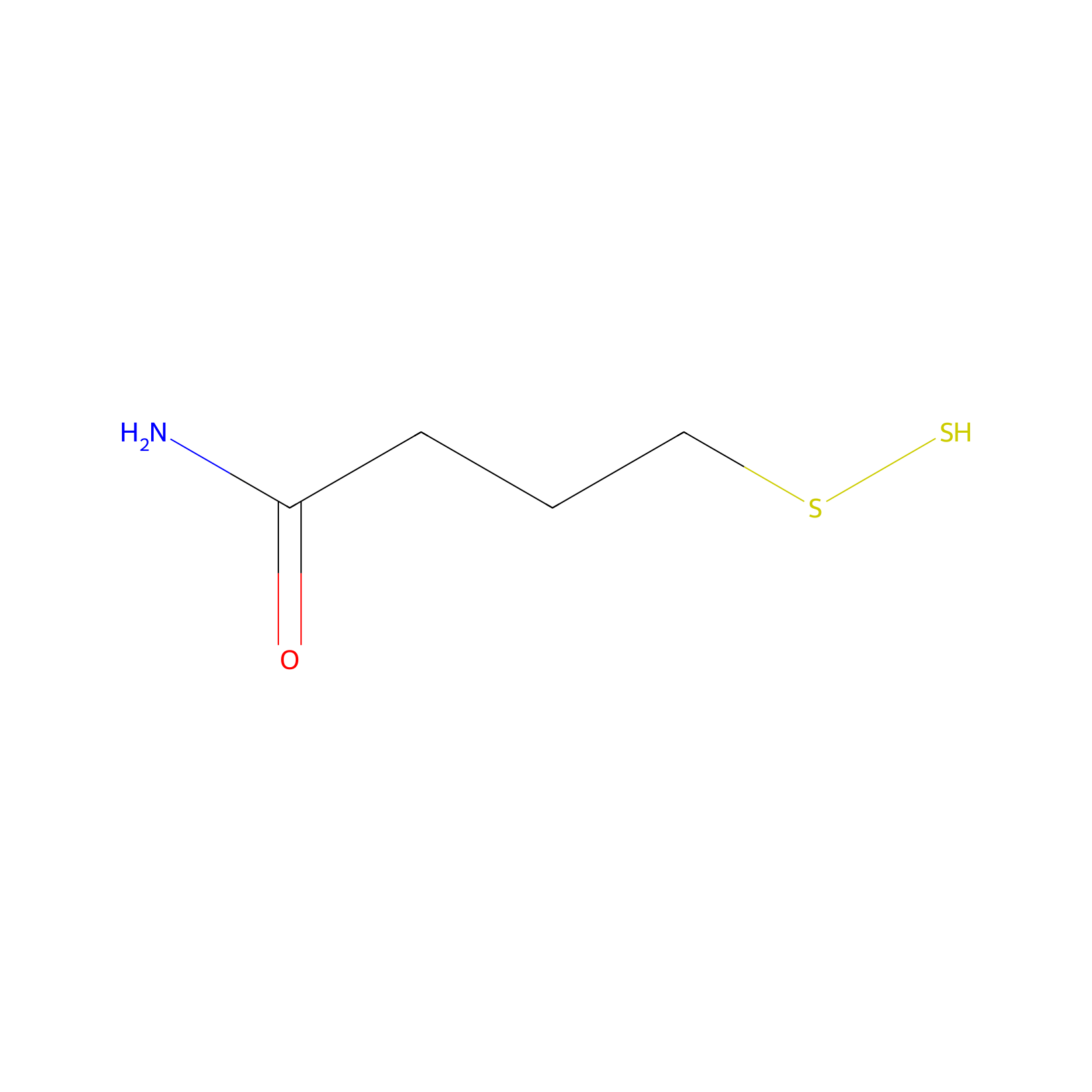
|
|||||
| Formula |
C4H9NOS2
|
|||||
| #Ro5 Violations (Lipinski): 0 | Molecular Weight (mw) | 151.3 | ||||
| Lipid-water partition coefficient (xlogp) | 0.2 | |||||
| Hydrogen Bond Donor Count (hbonddonor) | 2 | |||||
| Hydrogen Bond Acceptor Count (hbondacc) | 3 | |||||
| Rotatable Bond Count (rotbonds) | 4 | |||||
| PubChem CID | ||||||
| Canonical smiles |
C(CC(=O)N)CSS
|
|||||
| InChI |
InChI=1S/C4H9NOS2/c5-4(6)2-1-3-8-7/h7H,1-3H2,(H2,5,6)
|
|||||
| InChIKey |
AUDHCWSPRRYIGS-UHFFFAOYSA-N
|
|||||
| IUPAC Name |
4-(disulfanyl)butanamide
|
|||||
Each Peptide-drug Conjugate Related to This Linker
Full Information of The Activity Data of The PDC(s) Related to This Linker
(DOX-S)2-S-Pep [Investigative]
Discovered Using Cell Line-derived Xenograft Model
| Experiment 1 Reporting the Activity Data of This PDC | [1] | ||||
| Indication | Liver cancer | ||||
| Efficacy Data | Tumor volume |
1500 mm3
|
|||
| Administration Time | 24 day | ||||
| Administration Dosage | 6 mg/kg | ||||
| MOA of PDC |
Meanwhile, the thiol groups on Pep were used to covalently link it with a doxorubicin derivative (DOX-SH) and form PDC to kill tumors and inhibit tumor metastasis. Two doxorubicin molecules were linked to one Pep, and the drug loading was as high as 50.17%. The functional PDC molecule was synthesized from DOX-SH and 2,2-dipyridyl disulfide activated peptide (Pep-S-S-Py), and the molecular weight is 2565.98. CSNs were prepared for the effective codelivery of MMPI Pep and DOX to the tumor site. The MPL shell displayed a negative surface charge for prolonged blood circulation, and the PDC core was able to aggregate in the tumor matrix and adhere to the cell membrane. PDC aggregation could constantly release the MMPI peptide and DOX via low concentration and long-term glutathione-mediated reduction conditions in the tumor matrix. DOX can effectively enter the tumor cell and kill them. Meanwhile, MMPI peptide adherence to the cell membrane was able to selectively inhibit the activity of the MMP2 and achieve the effect of inhibiting tumor metastasis. Our study suggests that CSNs are of great potential for treating metastatic tumors.
Click to Show/Hide
|
||||
| Description |
The antitumor activity of CSNs was assessed in BALB/c male nude mice inoculated with high metastatic HCCLM3-LUC cells. As can be seen from Figure 3A,B, DOX caused severe systemic toxicity and a significant weight loss (up to 24.38%) during treatment. In contrast, there was little change in body weight for PDC- and CSNs-treated mice. Tumor progression was greatly suppressed in the CSNs group, as observed by bioluminescence. Tumor tissue images collected on day 27 confirmed that nude mice treated with CSNs bore the smallest tumors.
Click to Show/Hide
|
||||
| In Vivo Model | BALB/c male nude mice inoculated with high metastatic HCCLM3-LUC cells. | ||||
| In Vitro Model | Adult hepatocellular carcinoma | HCCLM3-Luc cell | CVCL_6832 | ||
| Experiment 2 Reporting the Activity Data of This PDC | [1] | ||||
| Indication | Liver cancer | ||||
| Efficacy Data | Lung metastasis nodules inhibition rates |
81.82%
|
|||
| Administration Time | 5 week | ||||
| Administration Dosage | 6 mg/kg | ||||
| Evaluation Method | H&E stain assay | ||||
| MOA of PDC |
Meanwhile, the thiol groups on Pep were used to covalently link it with a doxorubicin derivative (DOX-SH) and form PDC to kill tumors and inhibit tumor metastasis. Two doxorubicin molecules were linked to one Pep, and the drug loading was as high as 50.17%. The functional PDC molecule was synthesized from DOX-SH and 2,2-dipyridyl disulfide activated peptide (Pep-S-S-Py), and the molecular weight is 2565.98. CSNs were prepared for the effective codelivery of MMPI Pep and DOX to the tumor site. The MPL shell displayed a negative surface charge for prolonged blood circulation, and the PDC core was able to aggregate in the tumor matrix and adhere to the cell membrane. PDC aggregation could constantly release the MMPI peptide and DOX via low concentration and long-term glutathione-mediated reduction conditions in the tumor matrix. DOX can effectively enter the tumor cell and kill them. Meanwhile, MMPI peptide adherence to the cell membrane was able to selectively inhibit the activity of the MMP2 and achieve the effect of inhibiting tumor metastasis. Our study suggests that CSNs are of great potential for treating metastatic tumors.
Click to Show/Hide
|
||||
| Description |
For the PDC- and CSN-treated groups, the number of lung metastasis nodules was decreased (inhibition rates of 81.82% and 93.18%, respectively), and smaller areas of tumor cells were observed in the H&E-stained images. Nude mice treated with DOX displayed 7.8 nodules, which was not significantly different from what was seen in the PBS group.
|
||||
| In Vivo Model | BALB/c male nude mice inoculated with high metastatic HCCLM3-LUC cells. | ||||
| In Vitro Model | Adult hepatocellular carcinoma | HCCLM3-Luc cell | CVCL_6832 | ||
Revealed Based on the Cell Line Data
| Experiment 1 Reporting the Activity Data of This PDC | [1] | ||||
| Indication | Liver cancer | ||||
| Efficacy Data | Inhibitory effect |
82.84 ± 2.22%
|
|||
| Evaluation Method | Wound healing and transwell assays | ||||
| MOA of PDC |
Meanwhile, the thiol groups on Pep were used to covalently link it with a doxorubicin derivative (DOX-SH) and form PDC to kill tumors and inhibit tumor metastasis. Two doxorubicin molecules were linked to one Pep, and the drug loading was as high as 50.17%. The functional PDC molecule was synthesized from DOX-SH and 2,2-dipyridyl disulfide activated peptide (Pep-S-S-Py), and the molecular weight is 2565.98. CSNs were prepared for the effective codelivery of MMPI Pep and DOX to the tumor site. The MPL shell displayed a negative surface charge for prolonged blood circulation, and the PDC core was able to aggregate in the tumor matrix and adhere to the cell membrane. PDC aggregation could constantly release the MMPI peptide and DOX via low concentration and long-term glutathione-mediated reduction conditions in the tumor matrix. DOX can effectively enter the tumor cell and kill them. Meanwhile, MMPI peptide adherence to the cell membrane was able to selectively inhibit the activity of the MMP2 and achieve the effect of inhibiting tumor metastasis. Our study suggests that CSNs are of great potential for treating metastatic tumors.
Click to Show/Hide
|
||||
| Description |
The tumor invasion ability was also inhibited under the action of Pep and the PDC. Pep and PDC treatment resulted in a strong inhibition of invasion ability of SMCC-7721 cells with rates of 79.93 ± 3.86% and 82.84 ± 2.22%, respectively. These results were used to guide follow-up in vivo studies.
|
||||
| In Vitro Model | Hepatocellular carcinoma | SMMC-7721 cell | CVCL_0534 | ||
| Experiment 2 Reporting the Activity Data of This PDC | [1] | ||||
| Indication | Liver cancer | ||||
| Efficacy Data | Half Maximal Effective Concentration (EC50) |
20.22 µM
|
|||
| Evaluation Method | MTT colorimetric assay | ||||
| MOA of PDC |
Meanwhile, the thiol groups on Pep were used to covalently link it with a doxorubicin derivative (DOX-SH) and form PDC to kill tumors and inhibit tumor metastasis. Two doxorubicin molecules were linked to one Pep, and the drug loading was as high as 50.17%. The functional PDC molecule was synthesized from DOX-SH and 2,2-dipyridyl disulfide activated peptide (Pep-S-S-Py), and the molecular weight is 2565.98. CSNs were prepared for the effective codelivery of MMPI Pep and DOX to the tumor site. The MPL shell displayed a negative surface charge for prolonged blood circulation, and the PDC core was able to aggregate in the tumor matrix and adhere to the cell membrane. PDC aggregation could constantly release the MMPI peptide and DOX via low concentration and long-term glutathione-mediated reduction conditions in the tumor matrix. DOX can effectively enter the tumor cell and kill them. Meanwhile, MMPI peptide adherence to the cell membrane was able to selectively inhibit the activity of the MMP2 and achieve the effect of inhibiting tumor metastasis. Our study suggests that CSNs are of great potential for treating metastatic tumors.
Click to Show/Hide
|
||||
| Description |
An MTT colorimetric assay was used to evaluate the in vitro cytotoxicity of materials on SMMC-7721 cells. Within the concentration range of 1.25-100 uM, DOX, DOX-SH, and PDC had a certain killing effect on liver cancer cells. The IC50 values of DOX, DOX-SH, and PDC were 17.62, 14.75, and 20.22 uM, respectively.
|
||||
| In Vitro Model | Hepatocellular carcinoma | SMMC-7721 cell | CVCL_0534 | ||
References