
Linker Information
General Information of This Linker
| Linker ID |
LIN00017
|
|||||
|---|---|---|---|---|---|---|
| Linker Name |
PEG2
|
|||||
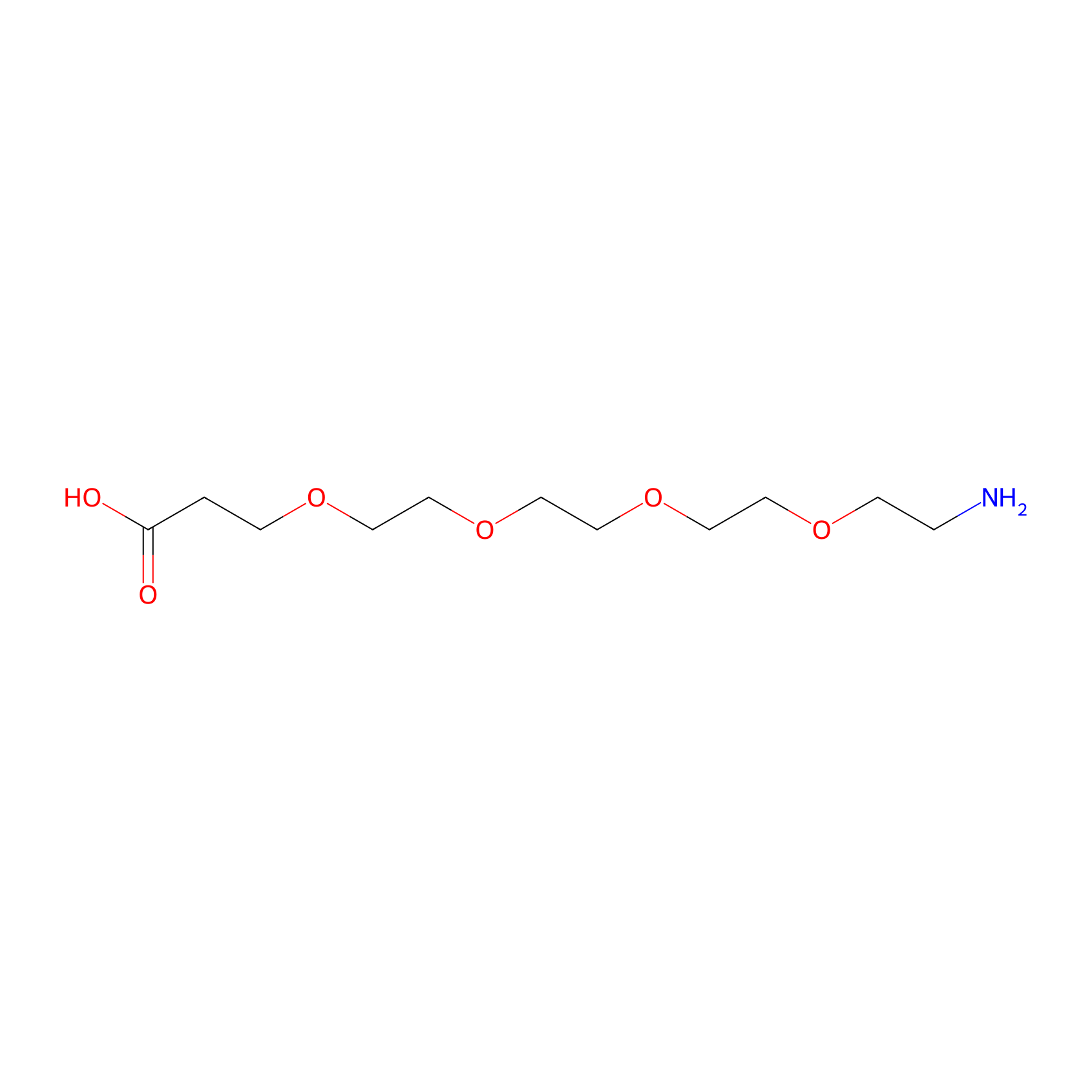
| Structure |
|
|||||
| Formula |
C11H23NO6
|
|||||
| #Ro5 Violations (Lipinski): 2 | Molecular Weight (mw) | 265.3 | ||||
| Lipid-water partition coefficient (xlogp) | -4.1 | |||||
| Hydrogen Bond Donor Count (hbonddonor) | 2 | |||||
| Hydrogen Bond Acceptor Count (hbondacc) | 7 | |||||
| Rotatable Bond Count (rotbonds) | 14 | |||||
| PubChem CID | ||||||
| Canonical smiles |
C(COCCOCCOCCOCCN)C(=O)O
|
|||||
| InChI |
InChI=1S/C11H23NO6/c12-2-4-16-6-8-18-10-9-17-7-5-15-3-1-11(13)14/h1-10,12H2,(H,13,14)
|
|||||
| InChIKey |
DKUZHSDZSMQOGQ-UHFFFAOYSA-N
|
|||||
| IUPAC Name |
3-[2-[2-[2-(2-aminoethoxy)ethoxy]ethoxy]ethoxy]propanoic acid
|
|||||
Each Peptide-drug Conjugate Related to This Linker
Full Information of The Activity Data of The PDC(s) Related to This Linker
99mTc-3PRGD2 [Phase 3]
Identified from the Human Clinical Data
| Experiment 1 Reporting the Activity Data of This PDC | [1] | ||||
| Indication | Primary malignant lung tumors | ||||
| Efficacy Data | Specificity |
88.24%
|
|||
| Patients Enrolled |
26 patients with primary malignant lung tumors.
|
||||
| Administration Time | 40-50 minutes | ||||
| Administration Dosage | 11.1 MBq (0.3 mCi)/kg | ||||
| MOA of PDC |
The integrin vβ3 receptor is a transmembrane heterodimer that mediates cell-cell and cell-extracellular matrix adhesions. It is ubiquitously present during the development and progression of malignant tumors and is closely associated with angiogenesis and metastasis. This receptor is highly expressed in proliferating tumor cells and activated endothelial cells, but is either not expressed or expressed at very low levels in normal endothelial cells, dormant vascular cells, and other normal cells, exhibiting a certain specificity. 99mtechnetium-three polyethylene glycol spacers-arginine-glycine-aspartic acid (99mTc-3PRGD2) is a single-photon emission computed tomography (SPECT) imaging agent that targets the integrin vβ33 receptor. Previous studies have confirmed the feasibility of 99mTc-3PRGD2 SPECT imaging for the diagnosis of lung cancer, breast cancer, esophageal cancer, and other malignant tumors and their related lymph node metastases. This study aimed to further investigate the performance of 99mTc-3PRGD2 SPECT/CT imaging in the diagnosis of lymph node metastasis in primary malignant lung tumors by comparing it with 18F-FDG PET/CT imaging.
Click to Show/Hide
|
||||
| Description |
Total 42 stations had metastatic lymph nodes and 136 stations had benign lymph nodes. The differences between metastatic and benign lymph nodes in the visual qualitative and semiquantitative analyses of 99mTc-3PRGD2 SPECT/CT and 18F-FDG PET/CT were statistically significant (all P < 0.001). The area under the receiver operating characteristic curve (AUC) in the semi-quantitative analysis of 99mTc-3PRGD2 SPECT/CT was 0.908 (95% confidence interval [CI], 0.851-0.966), and the sensitivity, specificity, positive predictive value, and negative predictive value were 0.86 (36/42), 0.88 (120/136), 0.69 (36/52), and 0.95 (120/126), respectively. Among the 26 patients (including two patients each with two lung tumors), 15 had pathologically confirmed lymph node metastasis. The difference between primary lung lesions in patients with and without lymph node metastasis was statistically significant only in the semi-quantitative analysis of 99mTc-3PRGD2 SPECT/CT (P = 0.007), with an AUC of 0.807 (95% CI, 0.641-0.974).
Click to Show/Hide
|
||||
| Experiment 2 Reporting the Activity Data of This PDC | [1] | ||||
| Indication | Primary malignant lung tumors | ||||
| Efficacy Data | Sensitivity |
85.71%
|
|||
| Patients Enrolled |
26 patients with primary malignant lung tumors.
|
||||
| Administration Time | 40-50 minutes | ||||
| Administration Dosage | 11.1 MBq (0.3 mCi)/kg | ||||
| MOA of PDC |
The integrin vβ3 receptor is a transmembrane heterodimer that mediates cell-cell and cell-extracellular matrix adhesions. It is ubiquitously present during the development and progression of malignant tumors and is closely associated with angiogenesis and metastasis. This receptor is highly expressed in proliferating tumor cells and activated endothelial cells, but is either not expressed or expressed at very low levels in normal endothelial cells, dormant vascular cells, and other normal cells, exhibiting a certain specificity. 99mtechnetium-three polyethylene glycol spacers-arginine-glycine-aspartic acid (99mTc-3PRGD2) is a single-photon emission computed tomography (SPECT) imaging agent that targets the integrin vβ33 receptor. Previous studies have confirmed the feasibility of 99mTc-3PRGD2 SPECT imaging for the diagnosis of lung cancer, breast cancer, esophageal cancer, and other malignant tumors and their related lymph node metastases. This study aimed to further investigate the performance of 99mTc-3PRGD2 SPECT/CT imaging in the diagnosis of lymph node metastasis in primary malignant lung tumors by comparing it with 18F-FDG PET/CT imaging.
Click to Show/Hide
|
||||
| Description |
Total 42 stations had metastatic lymph nodes and 136 stations had benign lymph nodes. The differences between metastatic and benign lymph nodes in the visual qualitative and semiquantitative analyses of 99mTc-3PRGD2 SPECT/CT and 18F-FDG PET/CT were statistically significant (all P < 0.001). The area under the receiver operating characteristic curve (AUC) in the semi-quantitative analysis of 99mTc-3PRGD2 SPECT/CT was 0.908 (95% confidence interval [CI], 0.851-0.966), and the sensitivity, specificity, positive predictive value, and negative predictive value were 0.86 (36/42), 0.88 (120/136), 0.69 (36/52), and 0.95 (120/126), respectively. Among the 26 patients (including two patients each with two lung tumors), 15 had pathologically confirmed lymph node metastasis. The difference between primary lung lesions in patients with and without lymph node metastasis was statistically significant only in the semi-quantitative analysis of 99mTc-3PRGD2 SPECT/CT (P = 0.007), with an AUC of 0.807 (95% CI, 0.641-0.974).
Click to Show/Hide
|
||||
| Experiment 3 Reporting the Activity Data of This PDC | [1] | ||||
| Indication | Primary malignant lung tumors | ||||
| Efficacy Data | Area under the curve (AUC) |
0.908
|
|||
| Patients Enrolled |
26 patients with primary malignant lung tumors.
|
||||
| Administration Time | 40-50 minutes | ||||
| Administration Dosage | 11.1 MBq (0.3 mCi)/kg | ||||
| MOA of PDC |
The integrin vβ3 receptor is a transmembrane heterodimer that mediates cell-cell and cell-extracellular matrix adhesions. It is ubiquitously present during the development and progression of malignant tumors and is closely associated with angiogenesis and metastasis. This receptor is highly expressed in proliferating tumor cells and activated endothelial cells, but is either not expressed or expressed at very low levels in normal endothelial cells, dormant vascular cells, and other normal cells, exhibiting a certain specificity. 99mtechnetium-three polyethylene glycol spacers-arginine-glycine-aspartic acid (99mTc-3PRGD2) is a single-photon emission computed tomography (SPECT) imaging agent that targets the integrin vβ33 receptor. Previous studies have confirmed the feasibility of 99mTc-3PRGD2 SPECT imaging for the diagnosis of lung cancer, breast cancer, esophageal cancer, and other malignant tumors and their related lymph node metastases. This study aimed to further investigate the performance of 99mTc-3PRGD2 SPECT/CT imaging in the diagnosis of lymph node metastasis in primary malignant lung tumors by comparing it with 18F-FDG PET/CT imaging.
Click to Show/Hide
|
||||
| Description |
Total 42 stations had metastatic lymph nodes and 136 stations had benign lymph nodes. The differences between metastatic and benign lymph nodes in the visual qualitative and semiquantitative analyses of 99mTc-3PRGD2 SPECT/CT and 18F-FDG PET/CT were statistically significant (all P < 0.001). The area under the receiver operating characteristic curve (AUC) in the semi-quantitative analysis of 99mTc-3PRGD2 SPECT/CT was 0.908 (95% confidence interval [CI], 0.851-0.966), and the sensitivity, specificity, positive predictive value, and negative predictive value were 0.86 (36/42), 0.88 (120/136), 0.69 (36/52), and 0.95 (120/126), respectively. Among the 26 patients (including two patients each with two lung tumors), 15 had pathologically confirmed lymph node metastasis. The difference between primary lung lesions in patients with and without lymph node metastasis was statistically significant only in the semi-quantitative analysis of 99mTc-3PRGD2 SPECT/CT (P = 0.007), with an AUC of 0.807 (95% CI, 0.641-0.974).
Click to Show/Hide
|
||||
References