
Peptide Information
General Information of This Peptide
| Peptide ID |
PEP00006
|
|||||
|---|---|---|---|---|---|---|
| Peptide Name |
CAAN-PTP-7
|
|||||
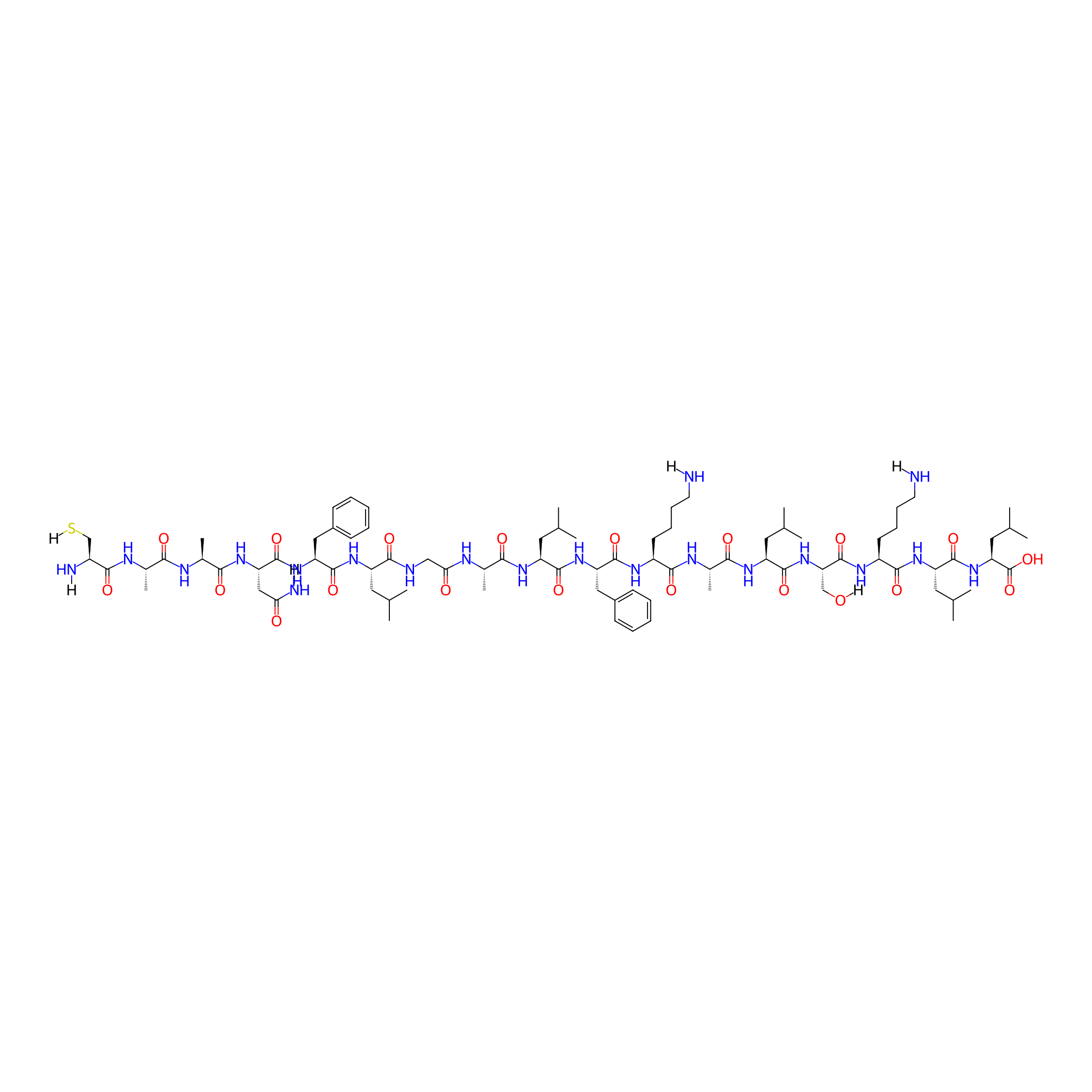
| Structure |
|
|||||
| Sequence |
CAANFLGALFKALSKLL
|
|||||
| Peptide Type |
Linear
|
|||||
| Receptor Name |
Cell membrane
|
|||||
| PDC Transmembrane Types | Cell-penetrating peptides (CPPs) | |||||
| Formula |
C84H138N20O20S
|
|||||
| Isosmiles |
[H]NCCCC[C@H](NC(=O)[C@H](Cc1ccccc1)NC(=O)[C@H](CC(C)C)NC(=O)[C@H](C)NC(=O)CNC(=O)[C@H](CC(C)C)NC(=O)[C@H](Cc1ccccc1)NC(=O)[C@H](CC(=O)N[H])NC(=O)[C@H](C)NC(=O)[C@H](C)NC(=O)[C@H](CS[H])N[H])C(=O)N[C@@H](C)C(=O)N[C@@H](CC(C)C)C(=O)N[C@@H](CO[H])C(=O)N[C@@H](CCCCN[H])C(=O)N[C@@H](CC(C)C)C(=O)N[C@@H](CC(C)C)C(=O)O
|
|||||
| InChI |
InChI=1S/C84H138N20O20S/c1-44(2)33-58(99-81(120)63(39-54-27-19-16-20-28-54)102-82(121)64(40-67(88)106)98-71(110)51(13)91-69(108)50(12)92-73(112)55(87)43-125)74(113)89-41-68(107)90-49(11)70(109)96-59(34-45(3)4)77(116)101-62(38-53-25-17-15-18-26-53)80(119)94-56(29-21-23-31-85)75(114)93-52(14)72(111)97-60(35-46(5)6)79(118)104-66(42-105)83(122)95-57(30-22-24-32-86)76(115)100-61(36-47(7)8)78(117)103-65(84(123)124)37-48(9)10/h15-20,25-28,44-52,55-66,105,125H,21-24,29-43,85-87H2,1-14H3,(H2,88,106)(H,89,113)(H,90,107)(H,91,108)(H,92,112)(H,93,114)(H,94,119)(H,95,122)(H,96,109)(H,97,111)(H,98,110)(H,99,120)(H,100,115)(H,101,116)(H,102,121)(H,103,117)(H,104,118)(H,123,124)/t49-,50-,51-,52-,55-,56-,57-,58-,59-,60-,61-,62-,63-,64-,65-,66-/m0/s1
|
|||||
| InChIKey |
IVZUILVPNIQSLS-FSXMPQNESA-N
|
|||||
| Pharmaceutical Properties |
Molecule Weight
|
1780.215
|
Polar area
|
644.28
|
||
|
Complexity
|
1779.011698
|
xlogp Value
|
-3.3625
|
|||
|
Heavy Count
|
125
|
Rot Bonds
|
64
|
|||
|
Hbond acc
|
23
|
Hbond Donor
|
23
|
|||
Each Peptide-drug Conjugate Related to This Peptide
Full Information of The Activity Data of The PDC(s) Related to This Peptide
mPEG-AAN-PTP-7-CDM-PTX [Investigative]
Revealed Based on the Cell Line Data
| Experiment 1 Reporting the Activity Data of This PDC | [1] | ||||
| Indication | Colon cancer | ||||
| Efficacy Data | Half Maximal Inhibitory Concentration (IC50) |
0.044 µg/mL
|
|||
| Administration Time | 72 h | ||||
| Evaluation Method | MTT assay | ||||
| MOA of PDC |
Herein, we present a lytic peptide PTP7-drug paclitaxel conjugate assembling nanoparticles (named PPP) that can sequentially respond to dual stimuli in the tumor microenvironment, which was designed for passive tumor-targeted delivery and on-demand release of a tumor lytic peptide (PTP-7) as well as a chemotherapeutic agent of paclitaxel (PTX). To achieve this, tumor lytic peptide PTP-7 was connected with polyethylene glycol by a peptide substrate of legumain to serve as hydrophobic segments of nanoparticles to protect the peptide from enzymatic degradation. After that, PTX was connected to the amino group of the polypeptide side chain through an acid-responsive chemical bond (2-propionic-3-methylmaleic anhydride, CDM). Therefore, the nanoparticle (PPP) collapsed when it encountered the weakly acidic tumor microenvironment where PTX molecules fell off, and further triggered the cleavage of the peptide substrate by legumain that is highly expressed in tumor stroma and tumor cell surface. Moreover, PPP presents improved stability, improved drug solubility, prolonged blood circulation and significant inhibition ability on tumor growth, which gives a reasonable strategy to accurately deliver small molecule drugs and active peptides simultaneously to tumor sites.
Click to Show/Hide
|
||||
| Description |
The cytotoxicity of PPP in MCF-7, HCT116, and 4T1 cells was investigated by MTT assay. The half maximal inhibitory concentration (IC50) of PPP NPs and free PTX was calculated to be 0.066 and 0.028 μg/mL on MCF-7 cells, 0.044 and 0.006 μg/mL on HCT116, 0.212 and 0.01 μg/mL on 4T1 cells which indicated a lower cell growth inhibition ability than free PTX. The result may be owing to the extremely stable property of PPP and low legumain expression in vitro leading to incomplete release of PTX and PTP-7 (Edgington et al., 2013). Moreover, the holographic microscopy studies showed the changes in cellular morphology of MCF-7 cells after treated with PPP for 3 h. Notably, PPP led to the morphological features of apoptosis such as shrinkage, losing contact with neighboring cells and floating relative to control.
Click to Show/Hide
|
||||
| In Vitro Model | Colon carcinoma | HCT 116 cell | CVCL_0291 | ||
| Experiment 2 Reporting the Activity Data of This PDC | [1] | ||||
| Indication | Breast cancer | ||||
| Efficacy Data | Half Maximal Inhibitory Concentration (IC50) |
0.066 µg/mL
|
|||
| Administration Time | 72 h | ||||
| Evaluation Method | MTT assay | ||||
| MOA of PDC |
Herein, we present a lytic peptide PTP7-drug paclitaxel conjugate assembling nanoparticles (named PPP) that can sequentially respond to dual stimuli in the tumor microenvironment, which was designed for passive tumor-targeted delivery and on-demand release of a tumor lytic peptide (PTP-7) as well as a chemotherapeutic agent of paclitaxel (PTX). To achieve this, tumor lytic peptide PTP-7 was connected with polyethylene glycol by a peptide substrate of legumain to serve as hydrophobic segments of nanoparticles to protect the peptide from enzymatic degradation. After that, PTX was connected to the amino group of the polypeptide side chain through an acid-responsive chemical bond (2-propionic-3-methylmaleic anhydride, CDM). Therefore, the nanoparticle (PPP) collapsed when it encountered the weakly acidic tumor microenvironment where PTX molecules fell off, and further triggered the cleavage of the peptide substrate by legumain that is highly expressed in tumor stroma and tumor cell surface. Moreover, PPP presents improved stability, improved drug solubility, prolonged blood circulation and significant inhibition ability on tumor growth, which gives a reasonable strategy to accurately deliver small molecule drugs and active peptides simultaneously to tumor sites.
Click to Show/Hide
|
||||
| Description |
The cytotoxicity of PPP in MCF-7, HCT116, and 4T1 cells was investigated by MTT assay. The half maximal inhibitory concentration (IC50) of PPP NPs and free PTX was calculated to be 0.066 and 0.028 μg/mL on MCF-7 cells, 0.044 and 0.006 μg/mL on HCT116, 0.212 and 0.01 μg/mL on 4T1 cells which indicated a lower cell growth inhibition ability than free PTX. The result may be owing to the extremely stable property of PPP and low legumain expression in vitro leading to incomplete release of PTX and PTP-7 (Edgington et al., 2013). Moreover, the holographic microscopy studies showed the changes in cellular morphology of MCF-7 cells after treated with PPP for 3 h. Notably, PPP led to the morphological features of apoptosis such as shrinkage, losing contact with neighboring cells and floating relative to control.
Click to Show/Hide
|
||||
| In Vitro Model | Invasive breast carcinoma | MCF-7 cell | CVCL_0031 | ||
| Experiment 3 Reporting the Activity Data of This PDC | [1] | ||||
| Indication | Breast cancer | ||||
| Efficacy Data | Half Maximal Inhibitory Concentration (IC50) |
0.212 µg/mL
|
|||
| Administration Time | 72 h | ||||
| Evaluation Method | MTT assay | ||||
| MOA of PDC |
Herein, we present a lytic peptide PTP7-drug paclitaxel conjugate assembling nanoparticles (named PPP) that can sequentially respond to dual stimuli in the tumor microenvironment, which was designed for passive tumor-targeted delivery and on-demand release of a tumor lytic peptide (PTP-7) as well as a chemotherapeutic agent of paclitaxel (PTX). To achieve this, tumor lytic peptide PTP-7 was connected with polyethylene glycol by a peptide substrate of legumain to serve as hydrophobic segments of nanoparticles to protect the peptide from enzymatic degradation. After that, PTX was connected to the amino group of the polypeptide side chain through an acid-responsive chemical bond (2-propionic-3-methylmaleic anhydride, CDM). Therefore, the nanoparticle (PPP) collapsed when it encountered the weakly acidic tumor microenvironment where PTX molecules fell off, and further triggered the cleavage of the peptide substrate by legumain that is highly expressed in tumor stroma and tumor cell surface. Moreover, PPP presents improved stability, improved drug solubility, prolonged blood circulation and significant inhibition ability on tumor growth, which gives a reasonable strategy to accurately deliver small molecule drugs and active peptides simultaneously to tumor sites.
Click to Show/Hide
|
||||
| Description |
The cytotoxicity of PPP in MCF-7, HCT116, and 4T1 cells was investigated by MTT assay. The half maximal inhibitory concentration (IC50) of PPP NPs and free PTX was calculated to be 0.066 and 0.028 μg/mL on MCF-7 cells, 0.044 and 0.006 μg/mL on HCT116, 0.212 and 0.01 μg/mL on 4T1 cells which indicated a lower cell growth inhibition ability than free PTX. The result may be owing to the extremely stable property of PPP and low legumain expression in vitro leading to incomplete release of PTX and PTP-7 (Edgington et al., 2013). Moreover, the holographic microscopy studies showed the changes in cellular morphology of MCF-7 cells after treated with PPP for 3 h. Notably, PPP led to the morphological features of apoptosis such as shrinkage, losing contact with neighboring cells and floating relative to control.
Click to Show/Hide
|
||||
| In Vitro Model | Mammary carcinoma | 4T1 cell | CVCL_0125 | ||
References