
Drug Information
General Information of This Drug
| Drug ID | DRG00014 | |||||
|---|---|---|---|---|---|---|
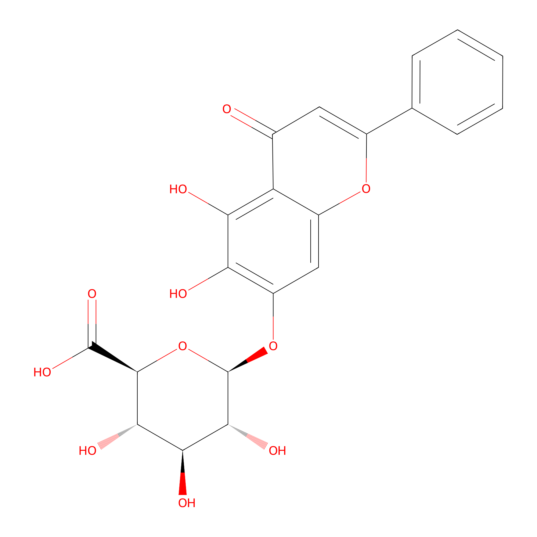
| Drug Name | Baicalin | |||||
| Synonyms |
Baicalin; 21967-41-9; Baicalein 7-O-glucuronide; 7-D-Glucuronic acid-5,6-dihydroxyflavone; Baicalein 7-glucuronide; CHEBI:2981; MFCD00134418; 347Q89U4M5; 5,6-dihydroxy-4-oxo-2-phenyl-4H-chromen-7-yl beta-D-glucopyranosiduronic acid; 7-D-glucuronic acid-5,6-dihydroxy-flavone; CHEMBL485818; UNII-347Q89U4M5; 5,6,7-trihydroxyflavone 7-O-beta-D-glucuronide; Baicalein 7-O-.beta.-D-glucuronide; beta-D-Glucopyranosiduronic acid, 5,6-dihydroxy-4-oxo-2-phenyl-4H-1-benzopyran-7-yl; (2S,3S,4S,5R,6S)-6-[(5,6-dihydroxy-4-oxo-2-phenyl-4H-chromen-7-yl)oxy]-3,4,5-trihydroxyoxane-2-carboxylic acid; BAICALEIN 7-O-BETA-D-GLUCURONIDE; 5,6-dihydroxy-4-oxo-2-phenyl-4H-1-benzopyran-7-yl beta-D-glucopyranosiduronic acid; 5,6-Dihydroxy-4-oxo-2-phenyl-4H-1-benzopyran-7-yl ; A-D-Glucopyranosiduronic Acid; BAICALEIN 7-O-GLUCURONIDE (USP-RS); BAICALEIN 7-O-GLUCURONIDE [USP-RS]; baikalin; Baicalein 7-O-; A-D-glucuronide; Baicalin,(S); (2S,3S,4S,5R,6R)-6-(5,6-dihydroxy-4-oxo-2-phenylchromen-7-yl)oxy-3,4,5-trihydroxyoxane-2-carboxylic acid; (2S,3S,4S,5R,6S)-6-(5,6-dihydroxy-4-oxo-2-phenyl-4H-chromen-7-yloxy)-3,4,5-trihydroxytetrahydro-2H-pyran-2-carboxylic acid; 0XE; ss-D-Glucopyranosiduronic acid, 5,6-dihydroxy-4-oxo-2-phenyl-4H-1-benzopyran-7-yl; Baicalin (6CI,7CI,8CI); Baicalein 7-O-glucuronide; Baicalein 7-O-ss-D-glucuronide; Baicalein 7-glucuronide; Baicalin, 95%; (2S,3S,4S,5R,6S)-6-((5,6-dihydroxy-4-oxo-2-phenyl-4H-chromen-7-yl)oxy)-3,4,5-trihydroxytetrahydro-2H-pyran-2-carboxylic acid; BAICALIN [VANDF]; Baicalein-7-D-glucuronide; BAICALIN [WHO-DD]; 31564-28-0; SCHEMBL285082; GTPL13076; TJN-151; DTXSID701346569; GLXC-10491; BAICALIN 7-B-D-GLUCURONIDE; Baicalin, >=99.0% (HPLC); HY-N0197; BDBM50242173; AKOS007930529; AKOS015955933; Baicalin 1000 microg/mL in Methanol; AC-7990; AM84780; CCG-214128; CS-5302; Baicalein 7-beta-D-glucopyranosiduronate; compound 15 [PMID: 37666112]; NCGC00386028-03; (2S,3S,4S,5R,6S)-6-(5,6-DIHYDROXY-4-OXO-2-PHENYL-CHROMEN-7-YL)OXY-3,4,5-TRIHYDROXY-OXANE-2-CARBOXYLIC ACID; (2S,3S,4S,5R,6S)-6-(5,6-dihydroxy-4-oxo-2-phenyl-chromen-7-yl)oxy-3,4,5-trihydroxy-tetrahydropyran-2-carboxylic acid; (2S,3S,4S,5R,6S)-6-(5,6-dihydroxy-4-oxo-2-phenylchromen-7-yl)oxy-3,4,5-trihydroxyoxane-2-carboxylic acid; 1ST40031; AS-13226; PD132941; NS00097648; A815791; J-013512; Q-100275; Q2879368; BRD-K49962337-001-01-1; Baicalin, European Pharmacopoeia (EP) Reference Standard; 5,6,7-Trihydroxyflavone-7-O-.beta.-D-glucopyranosideuronic acid; (2S,3S,4S,5R,6R)-6-(5,6-dihydroxy-4-oxo-2-phenyl-chromen-7-yl)oxy-3,4,5-trihydroxy-tetrahydropyran-2-carboxylic acid; (2S,3S,4S,5R,6S)-6-(5,6-dihydroxy-4-oxo-2-phenyl-4H-chromen-7-yloxy)-3,4,5-trihydroxy-tetrahydro-2H-pyran-2-carboxylic acid; (2S,3S,4S,5R,6S)-6-(5,6-dihydroxy-4-oxo-2-phenylchromen-7-yl)oxy-3,4,5-trihydroxyoxane-2-carboxylicacid; .BETA.-D-GLUCOPYRANOSIDURONIC ACID, 5,6-DIHYDROXY-4-OXO-2-PHENYL-4H-1-BENZOPYRAN-7-YL
Click to Show/Hide
|
|||||
| Structure |
|
|||||
| Formula |
C21H18O11
|
|||||
| #Ro5 Violations (Lipinski): 2 | Molecular Weight (mw) | 446.4 | ||||
| Lipid-water partition coefficient (xlogp) | 1.1 | |||||
| Hydrogen Bond Donor Count (hbonddonor) | 6 | |||||
| Hydrogen Bond Acceptor Count (hbondacc) | 11 | |||||
| Rotatable Bond Count (rotbonds) | 4 | |||||
| PubChem CID | ||||||
| Canonical smiles |
C1=CC=C(C=C1)C2=CC(=O)C3=C(C(=C(C=C3O2)OC4C(C(C(C(O4)C(=O)O)O)O)O)O)O
|
|||||
| InChI |
InChI=1S/C21H18O11/c22-9-6-10(8-4-2-1-3-5-8)30-11-7-12(14(23)15(24)13(9)11)31-21-18(27)16(25)17(26)19(32-21)20(28)29/h1-7,16-19,21,23-27H,(H,28,29)/t16-,17-,18+,19-,21+/m0/s1
|
|||||
| InChIKey |
IKIIZLYTISPENI-ZFORQUDYSA-N
|
|||||
| IUPAC Name |
(2S,3S,4S,5R,6S)-6-(5,6-dihydroxy-4-oxo-2-phenylchromen-7-yl)oxy-3,4,5-trihydroxyoxane-2-carboxylic acid
|
|||||
The activity data of This Drug
| Standard Type | Value | Administration times | Cell line | Cell line ID | Ref. | |
|---|---|---|---|---|---|---|
| Fe2+ decrease ratio | 25% | 24 h | H9c2 cell | CVCL_0286 | [1] | |
| GPX4/GAPDH increase ratio | 70% | 24 h | H9c2 cell | CVCL_0286 | [1] | |
| GSH/GSSG increase ratio | 40% | 24 h | H9c2 cell | CVCL_0286 | [1] | |
| MDA increase rate | 30% | 24 h | H9c2 cell | CVCL_0286 | [1] | |
| SLC7A11/GAPDH increase rate | 41% | 24 h | H9c2 cell | CVCL_0286 | [1] | |
| Half Maximal Inhibitory Concentration (IC50) | 72 uM | N.A. | H9 cell | CVCL_1240 | [2] | |
Full Information of The Activity Data of The PDC(s) Related to This Drug
BA-NFs [Investigative]
Revealed Based on the Cell Line Data
| Experiment 1 Reporting the Activity Data of This PDC | [1] | ||||
| Indication | DOX-induced cardiomyopathy | ||||
| Efficacy Data | SLC7A11/GAPDH increase rate | 83% | |||
| Evaluation Method | Western blotting assay | ||||
| Administration Time | 24 h | ||||
| Administration Dosage | 100 µM | ||||
| MOA of PDC |
Based on the above background, we designed and synthesized supramolecular self-assembled nanofibers with AT1R-specific targeting moieties (Baicalin-FFYEEG-ARVYIHPF, BA-NFs). Baicalin (BA), with its capacity for ROS scavenging and ferroptosis inhibition, constitutes an ideal candidate for ameliorating DIC. BA was conjugated with the self-assembling peptide FF and targeting peptide ARVYIHPF to form a specific nanostructure to improve its aqueous solubility and incorporate a targeting functionality, thereby addressing its inherent limitations. Our results indicated BA-NFs can mitigate doxorubicin-induced cardiomyocyte ferroptosis and cardiac dysfunction. This remarkable therapeutic effect is primarily attributable to the targeting proficiency of BA-NFs for injured cardiomyocytes, culminating in superoxide and ROS scavenging, reduced iron deposition, alleviated lipid peroxidation, and enhanced SLC7A11 and GPX4 expression. Therefore, we propose that BA-NFs proffers an efficacious targeted drug delivery strategy for the amelioration of DIC.
Click to Show/Hide
|
||||
| Description |
The accumulation of iron is a characteristic feature of ferroptosis. Intracellular iron, especially labile ferrous iron, can react with oxidants to generate cytotoxic hydroxyl radicals via Fenton reactions, thereby promoting ferroptosis. FerroOrange staining revealed significantly increased intracellular Fe2+ levels in DOX-treated H9c2 cells compared to controls, indicating DOX induced iron dyshomeostasis, resulting in ferrous iron buildup and subsequent ferroptosis. BA-NFs alleviated the accumulation of intracellular ferrous ions, suppressing ferroptosis. Fe2+ accumulation may disrupt the balance between LPO and oxygen homeostasis. We observed that BA-NFs efficiently attenuated DOX-induced elevation in malondialdehyde (MDA), a lipid peroxidation end-product, while increased antioxidant glutathione (GSH) levels. These findings suggested that BA-NFs can prevent DOX-induced ferroptosis in cardiomyocytes. Furthermore, protein expression of SLC7A11 and GPX4, key components of the antioxidant defense system against ferroptosis, was examined. DOX significantly decreased SLC7A11 and GPX4 expression in H9c2 cells compared to controls, suggesting DOX disrupts cellular antioxidant systems leading to ferroptosis. BA and BA-NFs partially restored SLC7A11 and GPX4 expression versus DOX treatment, with BA-NFs exhibiting the most significant impact. Together, these results indicate BA-NFs specifically inhibits DOX-induced cardiomyocyte ferroptosis by reducing intracellular iron accumulation and enhancing antioxidant system gene expression. In addition, the cardiotoxic effects of DOX are mediated via excessive oxidative stress and induced mitochondrial damage, thereby activating intrinsic apoptosis as an additional pathway. Flow cytometric analysis using Annexin V-Fitc/PI staining revealed that BA-NFs potently attenuated DOX-induced cardiomyocyte apoptosis, with a more pronounced effect than BA alone. DOX enhanced the green fluorescent signal of cleaved caspase-3 (as a biomarker of apoptosis) in H9c2 cells, whereas BA-NFs significantly inhibited this upregulation. These results provided further evidence that BA-NFs serve as potential therapeutic approaches for DIC.
Click to Show/Hide
|
||||
| In Vitro Model | Normal | H9c2 cell | CVCL_0286 | ||
| Experiment 2 Reporting the Activity Data of This PDC | [1] | ||||
| Indication | DOX-induced cardiomyopathy | ||||
| Efficacy Data | MDA increase rate | 50% | |||
| Evaluation Method | Western blotting assay | ||||
| Administration Time | 24 h | ||||
| Administration Dosage | 100 µM | ||||
| MOA of PDC |
Based on the above background, we designed and synthesized supramolecular self-assembled nanofibers with AT1R-specific targeting moieties (Baicalin-FFYEEG-ARVYIHPF, BA-NFs). Baicalin (BA), with its capacity for ROS scavenging and ferroptosis inhibition, constitutes an ideal candidate for ameliorating DIC. BA was conjugated with the self-assembling peptide FF and targeting peptide ARVYIHPF to form a specific nanostructure to improve its aqueous solubility and incorporate a targeting functionality, thereby addressing its inherent limitations. Our results indicated BA-NFs can mitigate doxorubicin-induced cardiomyocyte ferroptosis and cardiac dysfunction. This remarkable therapeutic effect is primarily attributable to the targeting proficiency of BA-NFs for injured cardiomyocytes, culminating in superoxide and ROS scavenging, reduced iron deposition, alleviated lipid peroxidation, and enhanced SLC7A11 and GPX4 expression. Therefore, we propose that BA-NFs proffers an efficacious targeted drug delivery strategy for the amelioration of DIC.
Click to Show/Hide
|
||||
| Description |
The accumulation of iron is a characteristic feature of ferroptosis. Intracellular iron, especially labile ferrous iron, can react with oxidants to generate cytotoxic hydroxyl radicals via Fenton reactions, thereby promoting ferroptosis. FerroOrange staining revealed significantly increased intracellular Fe2+ levels in DOX-treated H9c2 cells compared to controls, indicating DOX induced iron dyshomeostasis, resulting in ferrous iron buildup and subsequent ferroptosis. BA-NFs alleviated the accumulation of intracellular ferrous ions, suppressing ferroptosis. Fe2+ accumulation may disrupt the balance between LPO and oxygen homeostasis. We observed that BA-NFs efficiently attenuated DOX-induced elevation in malondialdehyde (MDA), a lipid peroxidation end-product, while increased antioxidant glutathione (GSH) levels. These findings suggested that BA-NFs can prevent DOX-induced ferroptosis in cardiomyocytes. Furthermore, protein expression of SLC7A11 and GPX4, key components of the antioxidant defense system against ferroptosis, was examined. DOX significantly decreased SLC7A11 and GPX4 expression in H9c2 cells compared to controls, suggesting DOX disrupts cellular antioxidant systems leading to ferroptosis. BA and BA-NFs partially restored SLC7A11 and GPX4 expression versus DOX treatment, with BA-NFs exhibiting the most significant impact. Together, these results indicate BA-NFs specifically inhibits DOX-induced cardiomyocyte ferroptosis by reducing intracellular iron accumulation and enhancing antioxidant system gene expression. In addition, the cardiotoxic effects of DOX are mediated via excessive oxidative stress and induced mitochondrial damage, thereby activating intrinsic apoptosis as an additional pathway. Flow cytometric analysis using Annexin V-Fitc/PI staining revealed that BA-NFs potently attenuated DOX-induced cardiomyocyte apoptosis, with a more pronounced effect than BA alone. DOX enhanced the green fluorescent signal of cleaved caspase-3 (as a biomarker of apoptosis) in H9c2 cells, whereas BA-NFs significantly inhibited this upregulation. These results provided further evidence that BA-NFs serve as potential therapeutic approaches for DIC.
Click to Show/Hide
|
||||
| In Vitro Model | Normal | H9c2 cell | CVCL_0286 | ||
| Experiment 3 Reporting the Activity Data of This PDC | [1] | ||||
| Indication | DOX-induced cardiomyopathy | ||||
| Efficacy Data | GSH/GSSG increase ratio | 85% | |||
| Evaluation Method | Western blotting assay | ||||
| Administration Time | 24 h | ||||
| Administration Dosage | 100 µM | ||||
| MOA of PDC |
Based on the above background, we designed and synthesized supramolecular self-assembled nanofibers with AT1R-specific targeting moieties (Baicalin-FFYEEG-ARVYIHPF, BA-NFs). Baicalin (BA), with its capacity for ROS scavenging and ferroptosis inhibition, constitutes an ideal candidate for ameliorating DIC. BA was conjugated with the self-assembling peptide FF and targeting peptide ARVYIHPF to form a specific nanostructure to improve its aqueous solubility and incorporate a targeting functionality, thereby addressing its inherent limitations. Our results indicated BA-NFs can mitigate doxorubicin-induced cardiomyocyte ferroptosis and cardiac dysfunction. This remarkable therapeutic effect is primarily attributable to the targeting proficiency of BA-NFs for injured cardiomyocytes, culminating in superoxide and ROS scavenging, reduced iron deposition, alleviated lipid peroxidation, and enhanced SLC7A11 and GPX4 expression. Therefore, we propose that BA-NFs proffers an efficacious targeted drug delivery strategy for the amelioration of DIC.
Click to Show/Hide
|
||||
| Description |
The accumulation of iron is a characteristic feature of ferroptosis. Intracellular iron, especially labile ferrous iron, can react with oxidants to generate cytotoxic hydroxyl radicals via Fenton reactions, thereby promoting ferroptosis. FerroOrange staining revealed significantly increased intracellular Fe2+ levels in DOX-treated H9c2 cells compared to controls, indicating DOX induced iron dyshomeostasis, resulting in ferrous iron buildup and subsequent ferroptosis. BA-NFs alleviated the accumulation of intracellular ferrous ions, suppressing ferroptosis. Fe2+ accumulation may disrupt the balance between LPO and oxygen homeostasis. We observed that BA-NFs efficiently attenuated DOX-induced elevation in malondialdehyde (MDA), a lipid peroxidation end-product, while increased antioxidant glutathione (GSH) levels. These findings suggested that BA-NFs can prevent DOX-induced ferroptosis in cardiomyocytes. Furthermore, protein expression of SLC7A11 and GPX4, key components of the antioxidant defense system against ferroptosis, was examined. DOX significantly decreased SLC7A11 and GPX4 expression in H9c2 cells compared to controls, suggesting DOX disrupts cellular antioxidant systems leading to ferroptosis. BA and BA-NFs partially restored SLC7A11 and GPX4 expression versus DOX treatment, with BA-NFs exhibiting the most significant impact. Together, these results indicate BA-NFs specifically inhibits DOX-induced cardiomyocyte ferroptosis by reducing intracellular iron accumulation and enhancing antioxidant system gene expression. In addition, the cardiotoxic effects of DOX are mediated via excessive oxidative stress and induced mitochondrial damage, thereby activating intrinsic apoptosis as an additional pathway. Flow cytometric analysis using Annexin V-Fitc/PI staining revealed that BA-NFs potently attenuated DOX-induced cardiomyocyte apoptosis, with a more pronounced effect than BA alone. DOX enhanced the green fluorescent signal of cleaved caspase-3 (as a biomarker of apoptosis) in H9c2 cells, whereas BA-NFs significantly inhibited this upregulation. These results provided further evidence that BA-NFs serve as potential therapeutic approaches for DIC.
Click to Show/Hide
|
||||
| In Vitro Model | Normal | H9c2 cell | CVCL_0286 | ||
| Experiment 4 Reporting the Activity Data of This PDC | [1] | ||||
| Indication | DOX-induced cardiomyopathy | ||||
| Efficacy Data | GPX4/GAPDH increase ratio | 120% | |||
| Evaluation Method | Western blotting assay | ||||
| Administration Time | 24 h | ||||
| Administration Dosage | 100 µM | ||||
| MOA of PDC |
Based on the above background, we designed and synthesized supramolecular self-assembled nanofibers with AT1R-specific targeting moieties (Baicalin-FFYEEG-ARVYIHPF, BA-NFs). Baicalin (BA), with its capacity for ROS scavenging and ferroptosis inhibition, constitutes an ideal candidate for ameliorating DIC. BA was conjugated with the self-assembling peptide FF and targeting peptide ARVYIHPF to form a specific nanostructure to improve its aqueous solubility and incorporate a targeting functionality, thereby addressing its inherent limitations. Our results indicated BA-NFs can mitigate doxorubicin-induced cardiomyocyte ferroptosis and cardiac dysfunction. This remarkable therapeutic effect is primarily attributable to the targeting proficiency of BA-NFs for injured cardiomyocytes, culminating in superoxide and ROS scavenging, reduced iron deposition, alleviated lipid peroxidation, and enhanced SLC7A11 and GPX4 expression. Therefore, we propose that BA-NFs proffers an efficacious targeted drug delivery strategy for the amelioration of DIC.
Click to Show/Hide
|
||||
| Description |
The accumulation of iron is a characteristic feature of ferroptosis. Intracellular iron, especially labile ferrous iron, can react with oxidants to generate cytotoxic hydroxyl radicals via Fenton reactions, thereby promoting ferroptosis. FerroOrange staining revealed significantly increased intracellular Fe2+ levels in DOX-treated H9c2 cells compared to controls, indicating DOX induced iron dyshomeostasis, resulting in ferrous iron buildup and subsequent ferroptosis. BA-NFs alleviated the accumulation of intracellular ferrous ions, suppressing ferroptosis. Fe2+ accumulation may disrupt the balance between LPO and oxygen homeostasis. We observed that BA-NFs efficiently attenuated DOX-induced elevation in malondialdehyde (MDA), a lipid peroxidation end-product, while increased antioxidant glutathione (GSH) levels. These findings suggested that BA-NFs can prevent DOX-induced ferroptosis in cardiomyocytes. Furthermore, protein expression of SLC7A11 and GPX4, key components of the antioxidant defense system against ferroptosis, was examined. DOX significantly decreased SLC7A11 and GPX4 expression in H9c2 cells compared to controls, suggesting DOX disrupts cellular antioxidant systems leading to ferroptosis. BA and BA-NFs partially restored SLC7A11 and GPX4 expression versus DOX treatment, with BA-NFs exhibiting the most significant impact. Together, these results indicate BA-NFs specifically inhibits DOX-induced cardiomyocyte ferroptosis by reducing intracellular iron accumulation and enhancing antioxidant system gene expression. In addition, the cardiotoxic effects of DOX are mediated via excessive oxidative stress and induced mitochondrial damage, thereby activating intrinsic apoptosis as an additional pathway. Flow cytometric analysis using Annexin V-Fitc/PI staining revealed that BA-NFs potently attenuated DOX-induced cardiomyocyte apoptosis, with a more pronounced effect than BA alone. DOX enhanced the green fluorescent signal of cleaved caspase-3 (as a biomarker of apoptosis) in H9c2 cells, whereas BA-NFs significantly inhibited this upregulation. These results provided further evidence that BA-NFs serve as potential therapeutic approaches for DIC.
Click to Show/Hide
|
||||
| In Vitro Model | Normal | H9c2 cell | CVCL_0286 | ||
| Experiment 5 Reporting the Activity Data of This PDC | [1] | ||||
| Indication | DOX-induced cardiomyopathy | ||||
| Efficacy Data | Fe2+ decrease ratio | 50% | |||
| Evaluation Method | Western blotting assay | ||||
| Administration Time | 24 h | ||||
| Administration Dosage | 100 µM | ||||
| MOA of PDC |
Based on the above background, we designed and synthesized supramolecular self-assembled nanofibers with AT1R-specific targeting moieties (Baicalin-FFYEEG-ARVYIHPF, BA-NFs). Baicalin (BA), with its capacity for ROS scavenging and ferroptosis inhibition, constitutes an ideal candidate for ameliorating DIC. BA was conjugated with the self-assembling peptide FF and targeting peptide ARVYIHPF to form a specific nanostructure to improve its aqueous solubility and incorporate a targeting functionality, thereby addressing its inherent limitations. Our results indicated BA-NFs can mitigate doxorubicin-induced cardiomyocyte ferroptosis and cardiac dysfunction. This remarkable therapeutic effect is primarily attributable to the targeting proficiency of BA-NFs for injured cardiomyocytes, culminating in superoxide and ROS scavenging, reduced iron deposition, alleviated lipid peroxidation, and enhanced SLC7A11 and GPX4 expression. Therefore, we propose that BA-NFs proffers an efficacious targeted drug delivery strategy for the amelioration of DIC.
Click to Show/Hide
|
||||
| Description |
The accumulation of iron is a characteristic feature of ferroptosis. Intracellular iron, especially labile ferrous iron, can react with oxidants to generate cytotoxic hydroxyl radicals via Fenton reactions, thereby promoting ferroptosis. FerroOrange staining revealed significantly increased intracellular Fe2+ levels in DOX-treated H9c2 cells compared to controls, indicating DOX induced iron dyshomeostasis, resulting in ferrous iron buildup and subsequent ferroptosis. BA-NFs alleviated the accumulation of intracellular ferrous ions, suppressing ferroptosis. Fe2+ accumulation may disrupt the balance between LPO and oxygen homeostasis. We observed that BA-NFs efficiently attenuated DOX-induced elevation in malondialdehyde (MDA), a lipid peroxidation end-product, while increased antioxidant glutathione (GSH) levels. These findings suggested that BA-NFs can prevent DOX-induced ferroptosis in cardiomyocytes. Furthermore, protein expression of SLC7A11 and GPX4, key components of the antioxidant defense system against ferroptosis, was examined. DOX significantly decreased SLC7A11 and GPX4 expression in H9c2 cells compared to controls, suggesting DOX disrupts cellular antioxidant systems leading to ferroptosis. BA and BA-NFs partially restored SLC7A11 and GPX4 expression versus DOX treatment, with BA-NFs exhibiting the most significant impact. Together, these results indicate BA-NFs specifically inhibits DOX-induced cardiomyocyte ferroptosis by reducing intracellular iron accumulation and enhancing antioxidant system gene expression. In addition, the cardiotoxic effects of DOX are mediated via excessive oxidative stress and induced mitochondrial damage, thereby activating intrinsic apoptosis as an additional pathway. Flow cytometric analysis using Annexin V-Fitc/PI staining revealed that BA-NFs potently attenuated DOX-induced cardiomyocyte apoptosis, with a more pronounced effect than BA alone. DOX enhanced the green fluorescent signal of cleaved caspase-3 (as a biomarker of apoptosis) in H9c2 cells, whereas BA-NFs significantly inhibited this upregulation. These results provided further evidence that BA-NFs serve as potential therapeutic approaches for DIC.
Click to Show/Hide
|
||||
| In Vitro Model | Normal | H9c2 cell | CVCL_0286 | ||
References