
Peptide-drug Conjugate Information
General Information of This Peptide-drug Conjugate (PDC)
| PDC ID |
PDC_02069
|
|||||
|---|---|---|---|---|---|---|
| PDC Name |
PDC-DOX2
|
|||||
| PDC Status |
Investigative
|
|||||
| Indication |
In total 1 Indication(s)
|
|||||
| Structure |
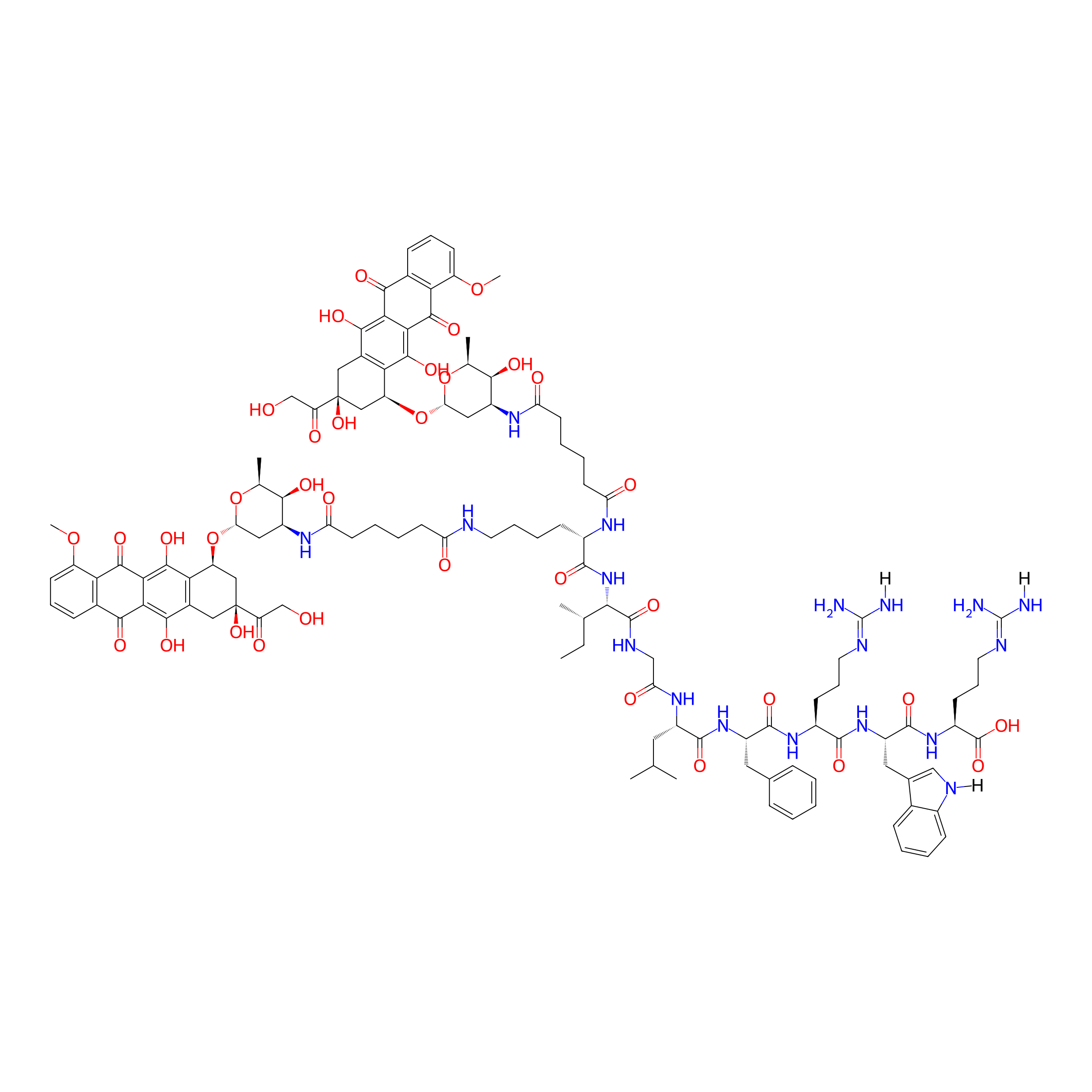
|
|||||
| Peptide Name |
KIGLFRWR
|
Peptide Info | ||||
| Drug Name |
Doxorubicin
|
Drug Info | ||||
| Therapeutic Target |
DNA topoisomerase 2-alpha (TOP2A)
|
Target Info | ||||
| Linker Name |
Adipic acid
|
Linker Info | ||||
| Peptide Modified Type |
Self-assembling
|
|||||
| Formula |
C118H152N18O35
|
|||||
| #Ro5 Violations (Lipinski): 4 | Molecular Weight | 2382.605 | ||||
| Lipid-water partition coefficient (xlogp) | 0.499 | |||||
| Hydrogen Bond Donor Count (hbonddonor) | 27 | |||||
| Hydrogen Bond Acceptor Count (hbondacc) | 36 | |||||
| Rotatable Bond Count (rotbonds) | 59 | |||||
Full List of Activity Data of This Peptide-drug Conjugate
Discovered Using Cell Line-derived Xenograft Model
| Experiment 1 Reporting the Activity Data of This PDC | [1] | ||||
| Indication | Hepatocellular carcinoma | ||||
| Efficacy Data | Body weight |
19g
|
|||
| Administration Time | 18 days | ||||
| Administration Dosage | 11 mg/kg | ||||
| MOA of PDC |
In this study, we designed and synthesized a novel peptide-drug conjugate (PDC-DOX2), in which two doxorubicin (DOX) molecules are covalently linked to a modified peptide with two carboxyl groups (Pep-AA). In neutral aqueous solution, PDC-DOX2 can self-assemble into stable spherical micelles due to hydrophilic-hydrophobic interactions. The sphere morphology can provide for the feasibility of intravenous injections of such peptide drug conjugates. PDC-DOX2 nanomicelles are stable spherical structures under neutral conditions, while they aggregate with decreased pH values. The pH value affected the assembly performance of PDC-DOX2 to a certain extent. With a decrease in pH (from a neutral to an acid environment), the morphology transforms from independent nanomicelles to slightly aggregated micelles and then to very aggregate micelles with diameters of nearly 3000 nm. The surfaces of PDC-DOX2 micelles were positively charged due to the lysine and arginine residues in the peptides. To avoid being engulfed by macrophages in plasma and prolong their blood circulation time, we further coated the positively charged micelles with a negatively charged natural polysaccharide shell, hyaluronic acid (HA), to form core-shell structure nanomedicine HA@PDC-DOX2. HA has various advantages, such as biodegradability, non-inflammatory, and non-immunogenicity. In addition, HA-coated nanomicelles allow for enhanced targeting in cancer therapy because HA can interact with overexpressed receptors in cancer cells, such as cluster determinant 44 (CD44), receptor for hyaluronic acid mediated motility (RHAMM) and intercellular adhesion molecule 1 (ICAM-1). Particularly, we found that the amount of HA influences the properties of HA@PDC-DOX2. The particle size of HA@PDC-DOX2 decreases with increasing HA content. The amount of HA can regulate the particle size, and HA@PDC-DOX2 become more stable in solution due to eliminating electrostatic repulsion of PDC-DOX2. The schematic mechanism of HA@PDC-DOX2 is shown in Scheme 1. First, PDC-DOX2 self-assembles into nanomicelles in neutral aqueous solution. Then, HA@PDC-DOX2 is constructed by negative HA shells and positively PDC-DOX2 cores. HA@PDC-DOX2 can deliver DOX into tumor sites via passive and active targeting effects. The core-shell structure HA@PDC-DOX2 nanomedicine showed better treatment effects on hepatocellular carcinoma, compared with PDC-DOX2 micelles and free DOX.
Click to Show/Hide
|
||||
| Description |
Body weight changes in all of the C57BL/6 mice in treatment groups, presented steady decreases.
|
||||
| In Vivo Model | H22 tumor-bearing C57BL/6 mice. | ||||
| Experiment 2 Reporting the Activity Data of This PDC | [1] | ||||
| Indication | Hepatocellular carcinoma | ||||
| Efficacy Data | Tumer volume |
580 mm3
|
|||
| Administration Time | 18 days | ||||
| Administration Dosage | 11 mg/kg | ||||
| MOA of PDC |
In this study, we designed and synthesized a novel peptide-drug conjugate (PDC-DOX2), in which two doxorubicin (DOX) molecules are covalently linked to a modified peptide with two carboxyl groups (Pep-AA). In neutral aqueous solution, PDC-DOX2 can self-assemble into stable spherical micelles due to hydrophilic-hydrophobic interactions. The sphere morphology can provide for the feasibility of intravenous injections of such peptide drug conjugates. PDC-DOX2 nanomicelles are stable spherical structures under neutral conditions, while they aggregate with decreased pH values. The pH value affected the assembly performance of PDC-DOX2 to a certain extent. With a decrease in pH (from a neutral to an acid environment), the morphology transforms from independent nanomicelles to slightly aggregated micelles and then to very aggregate micelles with diameters of nearly 3000 nm. The surfaces of PDC-DOX2 micelles were positively charged due to the lysine and arginine residues in the peptides. To avoid being engulfed by macrophages in plasma and prolong their blood circulation time, we further coated the positively charged micelles with a negatively charged natural polysaccharide shell, hyaluronic acid (HA), to form core-shell structure nanomedicine HA@PDC-DOX2. HA has various advantages, such as biodegradability, non-inflammatory, and non-immunogenicity. In addition, HA-coated nanomicelles allow for enhanced targeting in cancer therapy because HA can interact with overexpressed receptors in cancer cells, such as cluster determinant 44 (CD44), receptor for hyaluronic acid mediated motility (RHAMM) and intercellular adhesion molecule 1 (ICAM-1). Particularly, we found that the amount of HA influences the properties of HA@PDC-DOX2. The particle size of HA@PDC-DOX2 decreases with increasing HA content. The amount of HA can regulate the particle size, and HA@PDC-DOX2 become more stable in solution due to eliminating electrostatic repulsion of PDC-DOX2. The schematic mechanism of HA@PDC-DOX2 is shown in Scheme 1. First, PDC-DOX2 self-assembles into nanomicelles in neutral aqueous solution. Then, HA@PDC-DOX2 is constructed by negative HA shells and positively PDC-DOX2 cores. HA@PDC-DOX2 can deliver DOX into tumor sites via passive and active targeting effects. The core-shell structure HA@PDC-DOX2 nanomedicine showed better treatment effects on hepatocellular carcinoma, compared with PDC-DOX2 micelles and free DOX.
Click to Show/Hide
|
||||
| Description |
For the control group receiving PBS injections, the tumor volume expanded rapidly, whereas the tumor growth of the group receiving free DOX, PDC-DOX2, and HA@PDC-DOX2 could be suppressed to some degree. Among them, the inhibition in the HA@PDC-DOX2 group was the most obvious.
|
||||
| In Vivo Model | H22 tumor-bearing C57BL/6 mice. | ||||
Revealed Based on the Cell Line Data
| Experiment 1 Reporting the Activity Data of This PDC | [1] | ||||
| Indication | Hepatocellular carcinoma | ||||
| Efficacy Data | Cell viability |
38%
|
|||
| Administration Time | 4 h | ||||
| Administration Dosage | 50 μg/ml | ||||
| Evaluation Method | MTT assay | ||||
| MOA of PDC |
In this study, we designed and synthesized a novel peptide-drug conjugate (PDC-DOX2), in which two doxorubicin (DOX) molecules are covalently linked to a modified peptide with two carboxyl groups (Pep-AA). In neutral aqueous solution, PDC-DOX2 can self-assemble into stable spherical micelles due to hydrophilic-hydrophobic interactions. The sphere morphology can provide for the feasibility of intravenous injections of such peptide drug conjugates. PDC-DOX2 nanomicelles are stable spherical structures under neutral conditions, while they aggregate with decreased pH values. The pH value affected the assembly performance of PDC-DOX2 to a certain extent. With a decrease in pH (from a neutral to an acid environment), the morphology transforms from independent nanomicelles to slightly aggregated micelles and then to very aggregate micelles with diameters of nearly 3000 nm. The surfaces of PDC-DOX2 micelles were positively charged due to the lysine and arginine residues in the peptides. To avoid being engulfed by macrophages in plasma and prolong their blood circulation time, we further coated the positively charged micelles with a negatively charged natural polysaccharide shell, hyaluronic acid (HA), to form core-shell structure nanomedicine HA@PDC-DOX2. HA has various advantages, such as biodegradability, non-inflammatory, and non-immunogenicity. In addition, HA-coated nanomicelles allow for enhanced targeting in cancer therapy because HA can interact with overexpressed receptors in cancer cells, such as cluster determinant 44 (CD44), receptor for hyaluronic acid mediated motility (RHAMM) and intercellular adhesion molecule 1 (ICAM-1). Particularly, we found that the amount of HA influences the properties of HA@PDC-DOX2. The particle size of HA@PDC-DOX2 decreases with increasing HA content. The amount of HA can regulate the particle size, and HA@PDC-DOX2 become more stable in solution due to eliminating electrostatic repulsion of PDC-DOX2. The schematic mechanism of HA@PDC-DOX2 is shown in Scheme 1. First, PDC-DOX2 self-assembles into nanomicelles in neutral aqueous solution. Then, HA@PDC-DOX2 is constructed by negative HA shells and positively PDC-DOX2 cores. HA@PDC-DOX2 can deliver DOX into tumor sites via passive and active targeting effects. The core-shell structure HA@PDC-DOX2 nanomedicine showed better treatment effects on hepatocellular carcinoma, compared with PDC-DOX2 micelles and free DOX.
Click to Show/Hide
|
||||
| Description |
All of the samples inhibited tumor cell activity in a dose-dependent manner (0.1-50 μg/mL).The antitumor activity of PDC-DOX2 and HA@PDC-DOX2 was lower than that of free DOX (IC50 of DOX: 3.102 μg/mL, IC50 of PDC-DOX2: 7.449 μg/mL, IC50 of HA@PDC-DOX2: 24.05 μg/mL).
|
||||
| In Vitro Model | Hepatocellular carcinoma | SMMC-7721 cell | CVCL_0534 | ||
| Experiment 2 Reporting the Activity Data of This PDC | [1] | ||||
| Indication | Hepatocellular carcinoma | ||||
| Efficacy Data | Cell viability |
40%
|
|||
| Administration Time | 4 h | ||||
| Administration Dosage | 25 μg/ml | ||||
| Evaluation Method | MTT assay | ||||
| MOA of PDC |
In this study, we designed and synthesized a novel peptide-drug conjugate (PDC-DOX2), in which two doxorubicin (DOX) molecules are covalently linked to a modified peptide with two carboxyl groups (Pep-AA). In neutral aqueous solution, PDC-DOX2 can self-assemble into stable spherical micelles due to hydrophilic-hydrophobic interactions. The sphere morphology can provide for the feasibility of intravenous injections of such peptide drug conjugates. PDC-DOX2 nanomicelles are stable spherical structures under neutral conditions, while they aggregate with decreased pH values. The pH value affected the assembly performance of PDC-DOX2 to a certain extent. With a decrease in pH (from a neutral to an acid environment), the morphology transforms from independent nanomicelles to slightly aggregated micelles and then to very aggregate micelles with diameters of nearly 3000 nm. The surfaces of PDC-DOX2 micelles were positively charged due to the lysine and arginine residues in the peptides. To avoid being engulfed by macrophages in plasma and prolong their blood circulation time, we further coated the positively charged micelles with a negatively charged natural polysaccharide shell, hyaluronic acid (HA), to form core-shell structure nanomedicine HA@PDC-DOX2. HA has various advantages, such as biodegradability, non-inflammatory, and non-immunogenicity. In addition, HA-coated nanomicelles allow for enhanced targeting in cancer therapy because HA can interact with overexpressed receptors in cancer cells, such as cluster determinant 44 (CD44), receptor for hyaluronic acid mediated motility (RHAMM) and intercellular adhesion molecule 1 (ICAM-1). Particularly, we found that the amount of HA influences the properties of HA@PDC-DOX2. The particle size of HA@PDC-DOX2 decreases with increasing HA content. The amount of HA can regulate the particle size, and HA@PDC-DOX2 become more stable in solution due to eliminating electrostatic repulsion of PDC-DOX2. The schematic mechanism of HA@PDC-DOX2 is shown in Scheme 1. First, PDC-DOX2 self-assembles into nanomicelles in neutral aqueous solution. Then, HA@PDC-DOX2 is constructed by negative HA shells and positively PDC-DOX2 cores. HA@PDC-DOX2 can deliver DOX into tumor sites via passive and active targeting effects. The core-shell structure HA@PDC-DOX2 nanomedicine showed better treatment effects on hepatocellular carcinoma, compared with PDC-DOX2 micelles and free DOX.
Click to Show/Hide
|
||||
| Description |
All of the samples inhibited tumor cell activity in a dose-dependent manner (0.1-50 μg/mL).The antitumor activity of PDC-DOX2 and HA@PDC-DOX2 was lower than that of free DOX (IC50 of DOX: 3.102 μg/mL, IC50 of PDC-DOX2: 7.449 μg/mL, IC50 of HA@PDC-DOX2: 24.05 μg/mL).
|
||||
| In Vitro Model | Hepatocellular carcinoma | SMMC-7721 cell | CVCL_0534 | ||
| Experiment 3 Reporting the Activity Data of This PDC | [1] | ||||
| Indication | Hepatocellular carcinoma | ||||
| Efficacy Data | Cell viability |
48%
|
|||
| Administration Time | 4 h | ||||
| Administration Dosage | 10 μg/ml | ||||
| Evaluation Method | MTT assay | ||||
| MOA of PDC |
In this study, we designed and synthesized a novel peptide-drug conjugate (PDC-DOX2), in which two doxorubicin (DOX) molecules are covalently linked to a modified peptide with two carboxyl groups (Pep-AA). In neutral aqueous solution, PDC-DOX2 can self-assemble into stable spherical micelles due to hydrophilic-hydrophobic interactions. The sphere morphology can provide for the feasibility of intravenous injections of such peptide drug conjugates. PDC-DOX2 nanomicelles are stable spherical structures under neutral conditions, while they aggregate with decreased pH values. The pH value affected the assembly performance of PDC-DOX2 to a certain extent. With a decrease in pH (from a neutral to an acid environment), the morphology transforms from independent nanomicelles to slightly aggregated micelles and then to very aggregate micelles with diameters of nearly 3000 nm. The surfaces of PDC-DOX2 micelles were positively charged due to the lysine and arginine residues in the peptides. To avoid being engulfed by macrophages in plasma and prolong their blood circulation time, we further coated the positively charged micelles with a negatively charged natural polysaccharide shell, hyaluronic acid (HA), to form core-shell structure nanomedicine HA@PDC-DOX2. HA has various advantages, such as biodegradability, non-inflammatory, and non-immunogenicity. In addition, HA-coated nanomicelles allow for enhanced targeting in cancer therapy because HA can interact with overexpressed receptors in cancer cells, such as cluster determinant 44 (CD44), receptor for hyaluronic acid mediated motility (RHAMM) and intercellular adhesion molecule 1 (ICAM-1). Particularly, we found that the amount of HA influences the properties of HA@PDC-DOX2. The particle size of HA@PDC-DOX2 decreases with increasing HA content. The amount of HA can regulate the particle size, and HA@PDC-DOX2 become more stable in solution due to eliminating electrostatic repulsion of PDC-DOX2. The schematic mechanism of HA@PDC-DOX2 is shown in Scheme 1. First, PDC-DOX2 self-assembles into nanomicelles in neutral aqueous solution. Then, HA@PDC-DOX2 is constructed by negative HA shells and positively PDC-DOX2 cores. HA@PDC-DOX2 can deliver DOX into tumor sites via passive and active targeting effects. The core-shell structure HA@PDC-DOX2 nanomedicine showed better treatment effects on hepatocellular carcinoma, compared with PDC-DOX2 micelles and free DOX.
Click to Show/Hide
|
||||
| Description |
All of the samples inhibited tumor cell activity in a dose-dependent manner (0.1-50 μg/mL).The antitumor activity of PDC-DOX2 and HA@PDC-DOX2 was lower than that of free DOX (IC50 of DOX: 3.102 μg/mL, IC50 of PDC-DOX2: 7.449 μg/mL, IC50 of HA@PDC-DOX2: 24.05 μg/mL).
|
||||
| In Vitro Model | Hepatocellular carcinoma | SMMC-7721 cell | CVCL_0534 | ||
| Experiment 4 Reporting the Activity Data of This PDC | [1] | ||||
| Indication | Hepatocellular carcinoma | ||||
| Efficacy Data | Cell viability |
50%
|
|||
| Administration Time | 4 h | ||||
| Administration Dosage | 5 μg/ml | ||||
| Evaluation Method | MTT assay | ||||
| MOA of PDC |
In this study, we designed and synthesized a novel peptide-drug conjugate (PDC-DOX2), in which two doxorubicin (DOX) molecules are covalently linked to a modified peptide with two carboxyl groups (Pep-AA). In neutral aqueous solution, PDC-DOX2 can self-assemble into stable spherical micelles due to hydrophilic-hydrophobic interactions. The sphere morphology can provide for the feasibility of intravenous injections of such peptide drug conjugates. PDC-DOX2 nanomicelles are stable spherical structures under neutral conditions, while they aggregate with decreased pH values. The pH value affected the assembly performance of PDC-DOX2 to a certain extent. With a decrease in pH (from a neutral to an acid environment), the morphology transforms from independent nanomicelles to slightly aggregated micelles and then to very aggregate micelles with diameters of nearly 3000 nm. The surfaces of PDC-DOX2 micelles were positively charged due to the lysine and arginine residues in the peptides. To avoid being engulfed by macrophages in plasma and prolong their blood circulation time, we further coated the positively charged micelles with a negatively charged natural polysaccharide shell, hyaluronic acid (HA), to form core-shell structure nanomedicine HA@PDC-DOX2. HA has various advantages, such as biodegradability, non-inflammatory, and non-immunogenicity. In addition, HA-coated nanomicelles allow for enhanced targeting in cancer therapy because HA can interact with overexpressed receptors in cancer cells, such as cluster determinant 44 (CD44), receptor for hyaluronic acid mediated motility (RHAMM) and intercellular adhesion molecule 1 (ICAM-1). Particularly, we found that the amount of HA influences the properties of HA@PDC-DOX2. The particle size of HA@PDC-DOX2 decreases with increasing HA content. The amount of HA can regulate the particle size, and HA@PDC-DOX2 become more stable in solution due to eliminating electrostatic repulsion of PDC-DOX2. The schematic mechanism of HA@PDC-DOX2 is shown in Scheme 1. First, PDC-DOX2 self-assembles into nanomicelles in neutral aqueous solution. Then, HA@PDC-DOX2 is constructed by negative HA shells and positively PDC-DOX2 cores. HA@PDC-DOX2 can deliver DOX into tumor sites via passive and active targeting effects. The core-shell structure HA@PDC-DOX2 nanomedicine showed better treatment effects on hepatocellular carcinoma, compared with PDC-DOX2 micelles and free DOX.
Click to Show/Hide
|
||||
| Description |
All of the samples inhibited tumor cell activity in a dose-dependent manner (0.1-50 μg/mL).The antitumor activity of PDC-DOX2 and HA@PDC-DOX2 was lower than that of free DOX (IC50 of DOX: 3.102 μg/mL, IC50 of PDC-DOX2: 7.449 μg/mL, IC50 of HA@PDC-DOX2: 24.05 μg/mL).
|
||||
| In Vitro Model | Hepatocellular carcinoma | SMMC-7721 cell | CVCL_0534 | ||
| Experiment 5 Reporting the Activity Data of This PDC | [1] | ||||
| Indication | Hepatocellular carcinoma | ||||
| Efficacy Data | Cell viability |
58%
|
|||
| Administration Time | 4 h | ||||
| Administration Dosage | 2.5 μg/ml | ||||
| Evaluation Method | MTT assay | ||||
| MOA of PDC |
In this study, we designed and synthesized a novel peptide-drug conjugate (PDC-DOX2), in which two doxorubicin (DOX) molecules are covalently linked to a modified peptide with two carboxyl groups (Pep-AA). In neutral aqueous solution, PDC-DOX2 can self-assemble into stable spherical micelles due to hydrophilic-hydrophobic interactions. The sphere morphology can provide for the feasibility of intravenous injections of such peptide drug conjugates. PDC-DOX2 nanomicelles are stable spherical structures under neutral conditions, while they aggregate with decreased pH values. The pH value affected the assembly performance of PDC-DOX2 to a certain extent. With a decrease in pH (from a neutral to an acid environment), the morphology transforms from independent nanomicelles to slightly aggregated micelles and then to very aggregate micelles with diameters of nearly 3000 nm. The surfaces of PDC-DOX2 micelles were positively charged due to the lysine and arginine residues in the peptides. To avoid being engulfed by macrophages in plasma and prolong their blood circulation time, we further coated the positively charged micelles with a negatively charged natural polysaccharide shell, hyaluronic acid (HA), to form core-shell structure nanomedicine HA@PDC-DOX2. HA has various advantages, such as biodegradability, non-inflammatory, and non-immunogenicity. In addition, HA-coated nanomicelles allow for enhanced targeting in cancer therapy because HA can interact with overexpressed receptors in cancer cells, such as cluster determinant 44 (CD44), receptor for hyaluronic acid mediated motility (RHAMM) and intercellular adhesion molecule 1 (ICAM-1). Particularly, we found that the amount of HA influences the properties of HA@PDC-DOX2. The particle size of HA@PDC-DOX2 decreases with increasing HA content. The amount of HA can regulate the particle size, and HA@PDC-DOX2 become more stable in solution due to eliminating electrostatic repulsion of PDC-DOX2. The schematic mechanism of HA@PDC-DOX2 is shown in Scheme 1. First, PDC-DOX2 self-assembles into nanomicelles in neutral aqueous solution. Then, HA@PDC-DOX2 is constructed by negative HA shells and positively PDC-DOX2 cores. HA@PDC-DOX2 can deliver DOX into tumor sites via passive and active targeting effects. The core-shell structure HA@PDC-DOX2 nanomedicine showed better treatment effects on hepatocellular carcinoma, compared with PDC-DOX2 micelles and free DOX.
Click to Show/Hide
|
||||
| Description |
All of the samples inhibited tumor cell activity in a dose-dependent manner (0.1-50 μg/mL).The antitumor activity of PDC-DOX2 and HA@PDC-DOX2 was lower than that of free DOX (IC50 of DOX: 3.102 μg/mL, IC50 of PDC-DOX2: 7.449 μg/mL, IC50 of HA@PDC-DOX2: 24.05 μg/mL).
|
||||
| In Vitro Model | Hepatocellular carcinoma | SMMC-7721 cell | CVCL_0534 | ||
| Experiment 6 Reporting the Activity Data of This PDC | [1] | ||||
| Indication | Hepatocellular carcinoma | ||||
| Efficacy Data | Cell viability |
63%
|
|||
| Administration Time | 4 h | ||||
| Administration Dosage | 1 μg/ml | ||||
| Evaluation Method | MTT assay | ||||
| MOA of PDC |
In this study, we designed and synthesized a novel peptide-drug conjugate (PDC-DOX2), in which two doxorubicin (DOX) molecules are covalently linked to a modified peptide with two carboxyl groups (Pep-AA). In neutral aqueous solution, PDC-DOX2 can self-assemble into stable spherical micelles due to hydrophilic-hydrophobic interactions. The sphere morphology can provide for the feasibility of intravenous injections of such peptide drug conjugates. PDC-DOX2 nanomicelles are stable spherical structures under neutral conditions, while they aggregate with decreased pH values. The pH value affected the assembly performance of PDC-DOX2 to a certain extent. With a decrease in pH (from a neutral to an acid environment), the morphology transforms from independent nanomicelles to slightly aggregated micelles and then to very aggregate micelles with diameters of nearly 3000 nm. The surfaces of PDC-DOX2 micelles were positively charged due to the lysine and arginine residues in the peptides. To avoid being engulfed by macrophages in plasma and prolong their blood circulation time, we further coated the positively charged micelles with a negatively charged natural polysaccharide shell, hyaluronic acid (HA), to form core-shell structure nanomedicine HA@PDC-DOX2. HA has various advantages, such as biodegradability, non-inflammatory, and non-immunogenicity. In addition, HA-coated nanomicelles allow for enhanced targeting in cancer therapy because HA can interact with overexpressed receptors in cancer cells, such as cluster determinant 44 (CD44), receptor for hyaluronic acid mediated motility (RHAMM) and intercellular adhesion molecule 1 (ICAM-1). Particularly, we found that the amount of HA influences the properties of HA@PDC-DOX2. The particle size of HA@PDC-DOX2 decreases with increasing HA content. The amount of HA can regulate the particle size, and HA@PDC-DOX2 become more stable in solution due to eliminating electrostatic repulsion of PDC-DOX2. The schematic mechanism of HA@PDC-DOX2 is shown in Scheme 1. First, PDC-DOX2 self-assembles into nanomicelles in neutral aqueous solution. Then, HA@PDC-DOX2 is constructed by negative HA shells and positively PDC-DOX2 cores. HA@PDC-DOX2 can deliver DOX into tumor sites via passive and active targeting effects. The core-shell structure HA@PDC-DOX2 nanomedicine showed better treatment effects on hepatocellular carcinoma, compared with PDC-DOX2 micelles and free DOX.
Click to Show/Hide
|
||||
| Description |
All of the samples inhibited tumor cell activity in a dose-dependent manner (0.1-50 μg/mL).The antitumor activity of PDC-DOX2 and HA@PDC-DOX2 was lower than that of free DOX (IC50 of DOX: 3.102 μg/mL, IC50 of PDC-DOX2: 7.449 μg/mL, IC50 of HA@PDC-DOX2: 24.05 μg/mL).
|
||||
| In Vitro Model | Hepatocellular carcinoma | SMMC-7721 cell | CVCL_0534 | ||
| Experiment 7 Reporting the Activity Data of This PDC | [1] | ||||
| Indication | Hepatocellular carcinoma | ||||
| Efficacy Data | Cell viability |
84%
|
|||
| Administration Time | 4 h | ||||
| Administration Dosage | 0.1 μg/ml | ||||
| Evaluation Method | MTT assay | ||||
| MOA of PDC |
In this study, we designed and synthesized a novel peptide-drug conjugate (PDC-DOX2), in which two doxorubicin (DOX) molecules are covalently linked to a modified peptide with two carboxyl groups (Pep-AA). In neutral aqueous solution, PDC-DOX2 can self-assemble into stable spherical micelles due to hydrophilic-hydrophobic interactions. The sphere morphology can provide for the feasibility of intravenous injections of such peptide drug conjugates. PDC-DOX2 nanomicelles are stable spherical structures under neutral conditions, while they aggregate with decreased pH values. The pH value affected the assembly performance of PDC-DOX2 to a certain extent. With a decrease in pH (from a neutral to an acid environment), the morphology transforms from independent nanomicelles to slightly aggregated micelles and then to very aggregate micelles with diameters of nearly 3000 nm. The surfaces of PDC-DOX2 micelles were positively charged due to the lysine and arginine residues in the peptides. To avoid being engulfed by macrophages in plasma and prolong their blood circulation time, we further coated the positively charged micelles with a negatively charged natural polysaccharide shell, hyaluronic acid (HA), to form core-shell structure nanomedicine HA@PDC-DOX2. HA has various advantages, such as biodegradability, non-inflammatory, and non-immunogenicity. In addition, HA-coated nanomicelles allow for enhanced targeting in cancer therapy because HA can interact with overexpressed receptors in cancer cells, such as cluster determinant 44 (CD44), receptor for hyaluronic acid mediated motility (RHAMM) and intercellular adhesion molecule 1 (ICAM-1). Particularly, we found that the amount of HA influences the properties of HA@PDC-DOX2. The particle size of HA@PDC-DOX2 decreases with increasing HA content. The amount of HA can regulate the particle size, and HA@PDC-DOX2 become more stable in solution due to eliminating electrostatic repulsion of PDC-DOX2. The schematic mechanism of HA@PDC-DOX2 is shown in Scheme 1. First, PDC-DOX2 self-assembles into nanomicelles in neutral aqueous solution. Then, HA@PDC-DOX2 is constructed by negative HA shells and positively PDC-DOX2 cores. HA@PDC-DOX2 can deliver DOX into tumor sites via passive and active targeting effects. The core-shell structure HA@PDC-DOX2 nanomedicine showed better treatment effects on hepatocellular carcinoma, compared with PDC-DOX2 micelles and free DOX.
Click to Show/Hide
|
||||
| Description |
All of the samples inhibited tumor cell activity in a dose-dependent manner (0.1-50 μg/mL).The antitumor activity of PDC-DOX2 and HA@PDC-DOX2 was lower than that of free DOX (IC50 of DOX: 3.102 μg/mL, IC50 of PDC-DOX2: 7.449 μg/mL, IC50 of HA@PDC-DOX2: 24.05 μg/mL).
|
||||
| In Vitro Model | Hepatocellular carcinoma | SMMC-7721 cell | CVCL_0534 | ||
References