
Peptide Information
General Information of This Peptide
| Peptide ID |
PEP00060
|
|||||
|---|---|---|---|---|---|---|
| Peptide Name |
p4
|
|||||
| Structure |
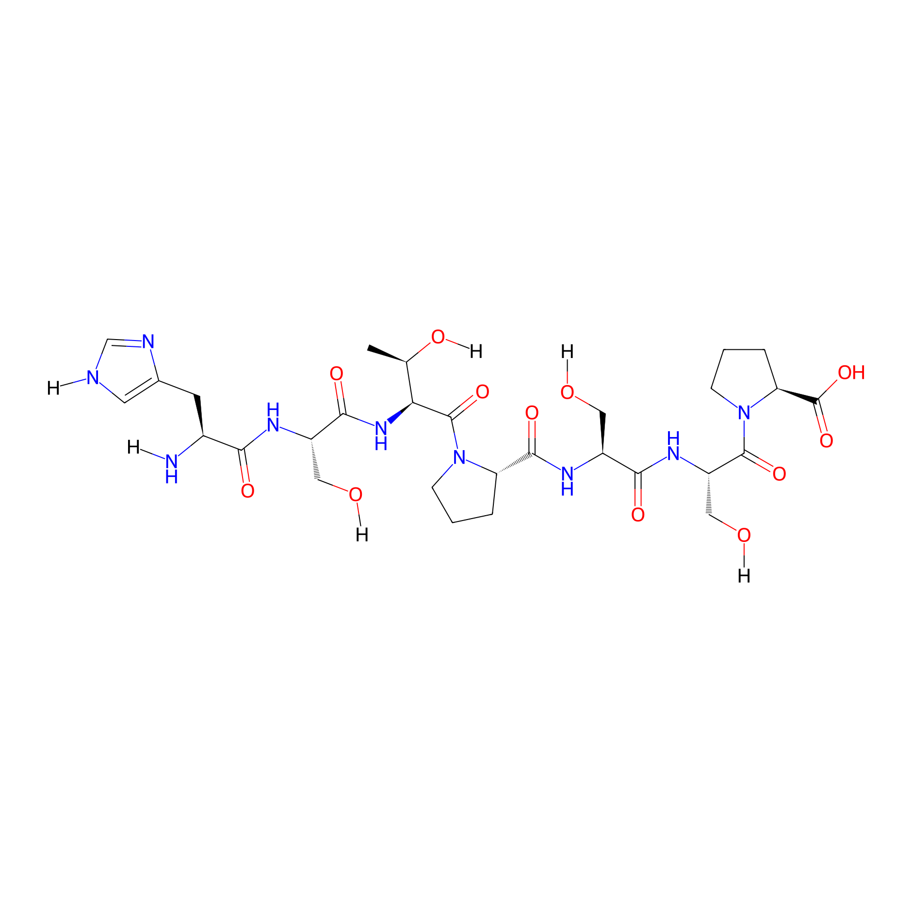
|
|||||
| Sequence |
HSTPSSP
|
|||||
| Peptide Type |
Linear
|
|||||
| PDC Transmembrane Types | Cell targeting peptides (CTPs) | |||||
| Formula |
C29H45N9O12
|
|||||
| Isosmiles |
[H]N[C@@H](Cc1cn([H])cn1)C(=O)N[C@@H](CO[H])C(=O)N[C@]([H])(C(=O)N1CCC[C@H]1C(=O)N[C@@H](CO[H])C(=O)N[C@@H](CO[H])C(=O)N1CCC[C@H]1C(=O)O)[C@@H](C)O[H]
|
|||||
| InChI |
InChI=1S/C29H45N9O12/c1-14(42)22(36-25(45)18(11-40)33-23(43)16(30)8-15-9-31-13-32-15)28(48)37-6-2-4-20(37)26(46)34-17(10-39)24(44)35-19(12-41)27(47)38-7-3-5-21(38)29(49)50/h9,13-14,16-22,39-42H,2-8,10-12,30H2,1H3,(H,31,32)(H,33,43)(H,34,46)(H,35,44)(H,36,45)(H,49,50)/t14-,16+,17+,18+,19+,20+,21+,22+/m1/s1
|
|||||
| InChIKey |
UGJZBVVDFNJCKQ-AJWYFOLQSA-N
|
|||||
| Pharmaceutical Properties |
Molecule Weight
|
711.73
|
Polar area
|
329.94
|
||
|
Complexity
|
711.3187679
|
xlogp Value
|
-6.357
|
|||
|
Heavy Count
|
50
|
Rot Bonds
|
22
|
|||
|
Hbond acc
|
13
|
Hbond Donor
|
11
|
|||
The Activity Data of This Peptide
| Peptide Activity Information 1 | [1] | |||||
| Binding rate | 0.82% | |||||
|---|---|---|---|---|---|---|
| Binding Affinity Assay |
To assess the binding capacity of the candidate peptides, peptide-FITC conjugates were incubated at 0, 4 or 8 ?M with 106 MOPC 315.BM cells at 37 C for 30 min.
|
|||||
| Experimental Condition | MOPC 315.BM cell | |||||
| Peptide Activity Information 2 | [1] | |||||
| Binding rate | 2.17% | |||||
| Binding Affinity Assay |
To assess the binding capacity of the candidate peptides, peptide-FITC conjugates were incubated at 0, 4 or 8 ?M with 106 PBMC cells at 37 C for 30 min.
|
|||||
| Experimental Condition | Mouse peripheral mononuclear cell PBMC | |||||
| Peptide Activity Information 3 | [1] | |||||
| Binding rate | 10.30% | |||||
| Binding Affinity Assay |
To assess the binding capacity of the candidate peptides, peptide-FITC conjugates were incubated at 0, 4 or 8 ?M with 106 NB4 cells at 37 C for 30 min.
|
|||||
| Experimental Condition | Human leukemic cell NB4 | |||||
| Peptide Activity Information 4 | [1] | |||||
| Binding rate | 13.80% | |||||
| Binding Affinity Assay |
To assess the binding capacity of the candidate peptides, peptide-FITC conjugates were incubated at 0, 4 or 8 ?M with 106 HL-60 cells at 37 C for 30 min.
|
|||||
| Experimental Condition | Human leukemic cell HL-60 | |||||
| Peptide Activity Information 5 | [1] | |||||
| Binding rate | 80.90% | |||||
| Binding Affinity Assay |
To assess the binding capacity of the candidate peptides, peptide-FITC conjugates were incubated at 0, 4 or 8 ?M with 106 A20 cells at 37 C for 30 min.
|
|||||
| Experimental Condition | A20 cell | |||||
Each Peptide-drug Conjugate Related to This Peptide
Full Information of The Activity Data of The PDC(s) Related to This Peptide
P4-Bend-PEG-AuNP [Investigative]
Revealed Based on the Cell Line Data
| Experiment 1 Reporting the Activity Data of This PDC | [1] | ||||
| Indication | Tumor | ||||
| Efficacy Data | A20 growth inhibition |
30.00%
|
|||
| Administration Time | 72 h | ||||
| Administration Dosage | 50 µM | ||||
| Evaluation Method | XTT assay | ||||
| Description |
All three P4-PDC-coated gold nanoparticles pre-incubated for 24 or 48 h induced statistically similar cytotoxicity in A20 to that induced by freshly prepared PDC4 and to coated particles without pre-incubation (the latter data not shown).
|
||||
| In Vitro Model | Mouse reticulum cell sarcoma | A20 cell | CVCL_1940 | ||
| Experiment 2 Reporting the Activity Data of This PDC | [1] | ||||
| Indication | Tumor | ||||
| Efficacy Data | A20 growth inhibition |
75.00%
|
|||
| Administration Time | 48 h | ||||
| Administration Dosage | 50 µM | ||||
| Evaluation Method | XTT assay | ||||
| Description |
All three P4-PDC-coated gold nanoparticles pre-incubated for 24 or 48 h induced statistically similar cytotoxicity in A20 to that induced by freshly prepared PDC4 and to coated particles without pre-incubation (the latter data not shown).
|
||||
| In Vitro Model | Mouse reticulum cell sarcoma | A20 cell | CVCL_1940 | ||
| Experiment 3 Reporting the Activity Data of This PDC | [1] | ||||
| Indication | Tumor | ||||
| Efficacy Data | A20 growth inhibition |
80.00%
|
|||
| Administration Time | 24 h | ||||
| Administration Dosage | 50 µM | ||||
| Evaluation Method | XTT assay | ||||
| Description |
All three P4-PDC-coated gold nanoparticles pre-incubated for 24 or 48 h induced statistically similar cytotoxicity in A20 to that induced by freshly prepared PDC4 and to coated particles without pre-incubation (the latter data not shown).
|
||||
| In Vitro Model | Mouse reticulum cell sarcoma | A20 cell | CVCL_1940 | ||
P4-Chlor-PEG-AuNP [Investigative]
Revealed Based on the Cell Line Data
| Experiment 1 Reporting the Activity Data of This PDC | [1] | ||||
| Indication | Tumor | ||||
| Efficacy Data | A20 growth inhibition |
40.00%
|
|||
| Administration Time | 72 h | ||||
| Administration Dosage | 50 µM | ||||
| Evaluation Method | XTT assay | ||||
| Description |
All three P4-PDC-coated gold nanoparticles pre-incubated for 24 or 48 h induced statistically similar cytotoxicity in A20 to that induced by freshly prepared PDC4 and to coated particles without pre-incubation (the latter data not shown).
|
||||
| In Vitro Model | Mouse reticulum cell sarcoma | A20 cell | CVCL_1940 | ||
| Experiment 2 Reporting the Activity Data of This PDC | [1] | ||||
| Indication | Tumor | ||||
| Efficacy Data | A20 growth inhibition |
72.00%
|
|||
| Administration Time | 48 h | ||||
| Administration Dosage | 50 µM | ||||
| Evaluation Method | XTT assay | ||||
| MOA of PDC |
In biological systems, antioxidants such as catalase, superoxide dismutase, glutathione peroxidase, and glutathione reductase are responsible for the elimination or reduction of the adverse effects of ROS, that is, they prevent or reduce ROS generation. Dietary antioxidants, such as vitamins E, A, and C, and anthocyanins and polyphenols have a role in the protection of cells against ROS damage.
Click to Show/Hide
|
||||
| Description |
All three P4-PDC-coated gold nanoparticles pre-incubated for 24 or 48 h induced statistically similar cytotoxicity in A20 to that induced by freshly prepared PDC4 and to coated particles without pre-incubation (the latter data not shown).
|
||||
| In Vitro Model | Mouse reticulum cell sarcoma | A20 cell | CVCL_1940 | ||
| Experiment 3 Reporting the Activity Data of This PDC | [1] | ||||
| Indication | Tumor | ||||
| Efficacy Data | A20 growth inhibition |
80.00%
|
|||
| Administration Time | 24 h | ||||
| Administration Dosage | 50 µM | ||||
| Evaluation Method | XTT assay | ||||
| Description |
All three P4-PDC-coated gold nanoparticles pre-incubated for 24 or 48 h induced statistically similar cytotoxicity in A20 to that induced by freshly prepared PDC4 and to coated particles without pre-incubation (the latter data not shown).
|
||||
| In Vitro Model | Mouse reticulum cell sarcoma | A20 cell | CVCL_1940 | ||
P4-Melph-PEG-AuNP [Investigative]
Revealed Based on the Cell Line Data
| Experiment 1 Reporting the Activity Data of This PDC | [1] | ||||
| Indication | Tumor | ||||
| Efficacy Data | A20 growth inhibition |
45.00%
|
|||
| Administration Time | 72 h | ||||
| Administration Dosage | 50 µM | ||||
| Evaluation Method | XTT assay | ||||
| Description |
All three P4-PDC-coated gold nanoparticles pre-incubated for 24 or 48 h induced statistically similar cytotoxicity in A20 to that induced by freshly prepared PDC4 and to coated particles without pre-incubation (the latter data not shown).
|
||||
| In Vitro Model | Mouse reticulum cell sarcoma | A20 cell | CVCL_1940 | ||
| Experiment 2 Reporting the Activity Data of This PDC | [1] | ||||
| Indication | Tumor | ||||
| Efficacy Data | A20 growth inhibition |
78.00%
|
|||
| Administration Time | 48 h | ||||
| Administration Dosage | 50 µM | ||||
| Evaluation Method | XTT assay | ||||
| MOA of PDC |
Vitamin C as a water-soluble vitamin is the reduced form of ascorbic acid. No significant adverse effect of taking high doses of vitamin C (over 2000 mg/day) has been reported due to the water-soluble feature of vitamin C. Vitamin C directly reacts with hydroxy, alkoxyl, and lipid peroxyl radicals and converts them to alcohol, water, and hydroperoxide lipid, respectively. It has been shown that taking vitamin C before radioiodine therapy can ameliorate the oxidative stress effect of radioiodine. The radioprotective effects of vitamin C are mainly due to its free radical scavenging activity.
Click to Show/Hide
|
||||
| Description |
All three P4-PDC-coated gold nanoparticles pre-incubated for 24 or 48 h induced statistically similar cytotoxicity in A20 to that induced by freshly prepared PDC4 and to coated particles without pre-incubation (the latter data not shown).
|
||||
| In Vitro Model | Mouse reticulum cell sarcoma | A20 cell | CVCL_1940 | ||
| Experiment 3 Reporting the Activity Data of This PDC | [1] | ||||
| Indication | Tumor | ||||
| Efficacy Data | A20 growth inhibition |
80.00%
|
|||
| Administration Time | 24 h | ||||
| Administration Dosage | 50 µM | ||||
| Evaluation Method | XTT assay | ||||
| Description |
All three P4-PDC-coated gold nanoparticles pre-incubated for 24 or 48 h induced statistically similar cytotoxicity in A20 to that induced by freshly prepared PDC4 and to coated particles without pre-incubation (the latter data not shown).
|
||||
| In Vitro Model | Mouse reticulum cell sarcoma | A20 cell | CVCL_1940 | ||
P4-chlorambucil [Investigative]
Revealed Based on the Cell Line Data
| Experiment 1 Reporting the Activity Data of This PDC | [1] | ||||
| Indication | Tumor | ||||
| Efficacy Data | A20 growth inhibition |
70.00%
|
|||
| Administration Time | 72 h | ||||
| Administration Dosage | 50 µM | ||||
| Evaluation Method | XTT assay | ||||
| Description |
Conjugation of the drugs to P4 affected their efficacy toward A20 cells. For chlorambucil and melphalan, conjugation reduced the cytotoxic effect and this was significant for chlorambucil at 25 μM (p = 0.0013). On the other hand, conjugation significantly improved the cytotoxic effect of bendamustine at 25 (p = 0.043) and 50 μM (p = 0.048). The efficacies of all P6-conjugates were significantly lower than those of P4-conjugates at concentrations above 10 μM.
Click to Show/Hide
|
||||
| In Vitro Model | Mouse reticulum cell sarcoma | A20 cell | CVCL_1940 | ||
P4-bendamustine [Investigative]
Revealed Based on the Cell Line Data
| Experiment 1 Reporting the Activity Data of This PDC | [1] | ||||
| Indication | Tumor | ||||
| Efficacy Data | A20 growth inhibition |
70.00%
|
|||
| Administration Time | 72 h | ||||
| Administration Dosage | 50 µM | ||||
| Evaluation Method | XTT assay | ||||
| Description |
Conjugation of the drugs to P4 affected their efficacy toward A20 cells. For chlorambucil and melphalan, conjugation reduced the cytotoxic effect and this was significant for chlorambucil at 25 μM (p = 0.0013). On the other hand, conjugation significantly improved the cytotoxic effect of bendamustine at 25 (p = 0.043) and 50 μM (p = 0.048). The efficacies of all P6-conjugates were significantly lower than those of P4-conjugates at concentrations above 10 μM.
Click to Show/Hide
|
||||
| In Vitro Model | Mouse reticulum cell sarcoma | A20 cell | CVCL_1940 | ||
P4-melphalan [Investigative]
Revealed Based on the Cell Line Data
| Experiment 1 Reporting the Activity Data of This PDC | [1] | ||||
| Indication | Tumor | ||||
| Efficacy Data | A20 growth inhibition |
75.00%
|
|||
| Administration Time | 72 h | ||||
| Administration Dosage | 50 µM | ||||
| Evaluation Method | XTT assay | ||||
| Description |
Conjugation of the drugs to P4 affected their efficacy toward A20 cells. For chlorambucil and melphalan, conjugation reduced the cytotoxic effect and this was significant for chlorambucil at 25 μM (p = 0.0013). On the other hand, conjugation significantly improved the cytotoxic effect of bendamustine at 25 (p = 0.043) and 50 μM (p = 0.048). The efficacies of all P6-conjugates were significantly lower than those of P4-conjugates at concentrations above 10 μM.
Click to Show/Hide
|
||||
| In Vitro Model | Mouse reticulum cell sarcoma | A20 cell | CVCL_1940 | ||
References