
Peptide-drug Conjugate Information
General Information of This Peptide-drug Conjugate (PDC)
| PDC ID |
PDC_02131
|
|||||
|---|---|---|---|---|---|---|
| PDC Name |
POSS-p18-4-Ce6(PPC)
|
|||||
| PDC Status |
Investigative
|
|||||
| Indication |
In total 1 Indication(s)
|
|||||
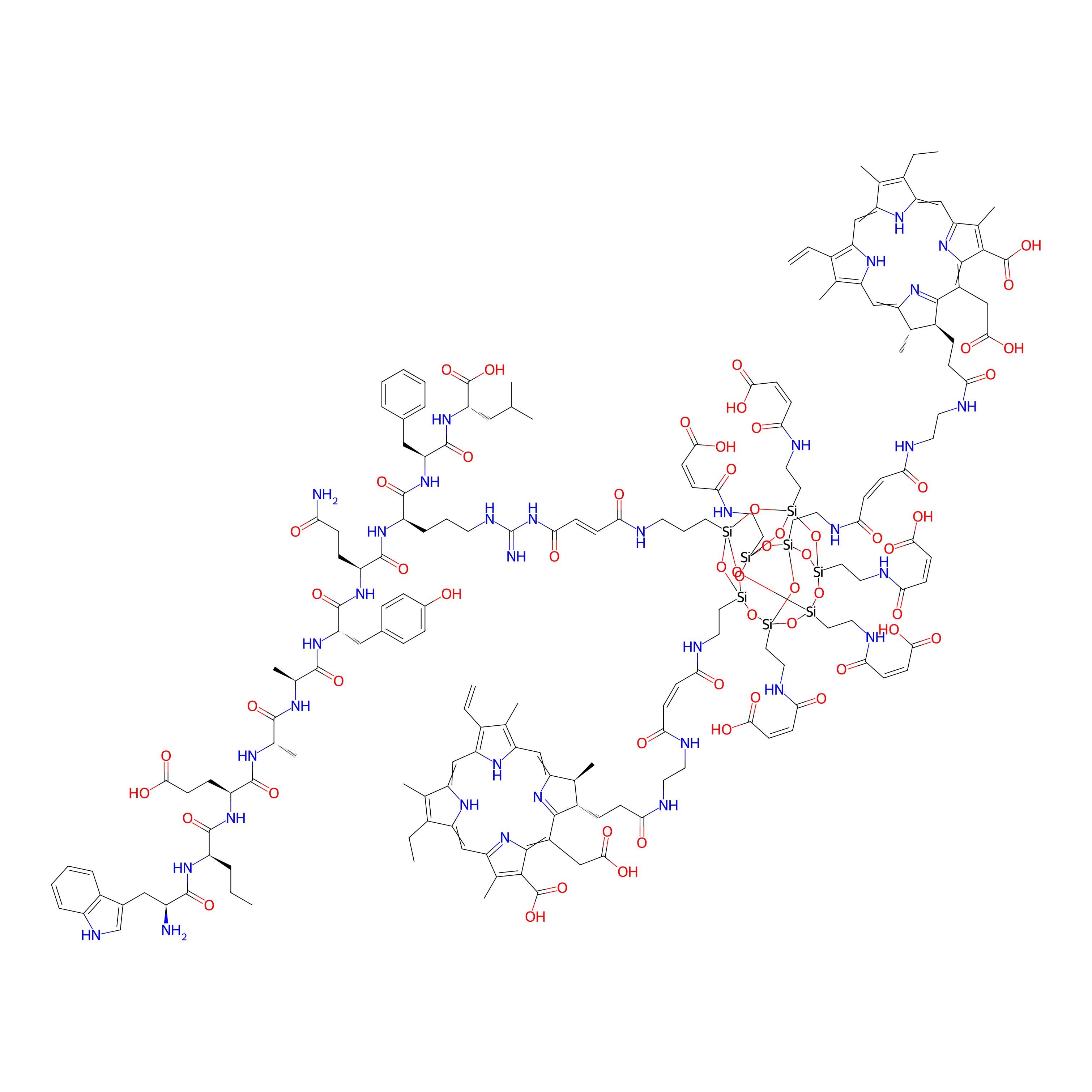
| Structure |
|
|||||
| Peptide Name |
Peptide 18-4
|
Peptide Info | ||||
| Receptor Name |
Keratin, type II cytoskeletal 1 (KRT1)
|
Receptor Info | ||||
| Drug Name |
Chlorin e6
|
Drug Info | ||||
| Linker Name |
POSS
|
Linker Info | ||||
| Formula |
C183H231N35O58Si8
|
|||||
| #Ro5 Violations (Lipinski): 4 | Molecular Weight | 4073.736 | ||||
| Lipid-water partition coefficient (xlogp) | 4.47015 | |||||
| Hydrogen Bond Donor Count (hbonddonor) | 43 | |||||
| Hydrogen Bond Acceptor Count (hbondacc) | 53 | |||||
| Rotatable Bond Count (rotbonds) | 104 | |||||
Full List of Activity Data of This Peptide-drug Conjugate
Revealed Based on the Cell Line Data
| Experiment 1 Reporting the Activity Data of This PDC | [1] | ||||
| Indication | Triple-negative breast cancer | ||||
| Efficacy Data | Cell viability |
10%
|
|||
| Administration Time | 24 h | ||||
| Evaluation Method | MTT assay | ||||
| MOA of PDC |
Polyhedral oligomeric silsesquioxane (POSS) molecules have a distinct nanostructure, consisting of an inner inorganic cage core of silicon and oxygen atoms and an outer shell of organic functional groups. The unique structure of POSS molecules containing reactive functionalities and their superior biocompatibility make them suitable drug-delivery carriers. In the present study, we prepared p 18-4/chlorin e6 (Ce6)-conjugated POSS (PPC) nanoparticles for improving the targeting ability of Ce6 to breast cancer cells. To fabricate PPC nanoparticles, p 18-4 was first covalently conjugated on OctaMaleamicacid POSS (OM-POSS), and subsequently, amino-terminated Ce6 was introduced. To verify the availability of the PPC nanoparticles for cancer therapy, their physicochemical properties, including morphology, chemical structure, and particle size, were systematically characterized. The cellular uptake, phototoxicity, and targeting ability of the PPC nanoparticles were assessed using a human breast cancer cell line MDA-MB-231. In addition, PPC nanoparticles was injected intravenously into tumor-bearing mice to evaluate in vivo biodistribution and PDT efficacy.
Click to Show/Hide
|
||||
| Description |
The internalization of nanoparticles in cancer cells is the foremost step in PDT. Targeting ligand decoration and reducing nanoparticle size have been intensively explored to enhance the intracellular uptake of nanoparticles. Therefore, the PPC nanoparticles were designed to offer the appropriate size and targeting ability for delivery of Ce6 to tumor tissues via active and passive targeting mechanisms. The cellular internalization of free Ce6 and PPC into various cancer cells, i.e., MDA-MB-231, MKN-28, and HeLa, was assessed by fluorescence-activated cell sorting (FACS) analysis using a flow cytometer. When compared with free Ce6, the higher cellular internalization of PPC was observed in all cancer cells tested. In particular, the fluorescence intensity due to the intracellular uptake of PPC in MDA-MB-231 cells increased more dramatically than in the other cancer cells.
Click to Show/Hide
|
||||
| In Vitro Model | Breast adenocarcinoma | MDA-MB-231 cell | CVCL_0062 | ||
References