
Peptide-drug Conjugate Information
General Information of This Peptide-drug Conjugate (PDC)
| PDC ID |
PDC_00347
|
|||||
|---|---|---|---|---|---|---|
| PDC Name |
Tesaglitazar-[F7, P34]-NPY
|
|||||
| PDC Status |
Investigative
|
|||||
| Indication |
In total 1 Indication(s)
|
|||||
| Structure |
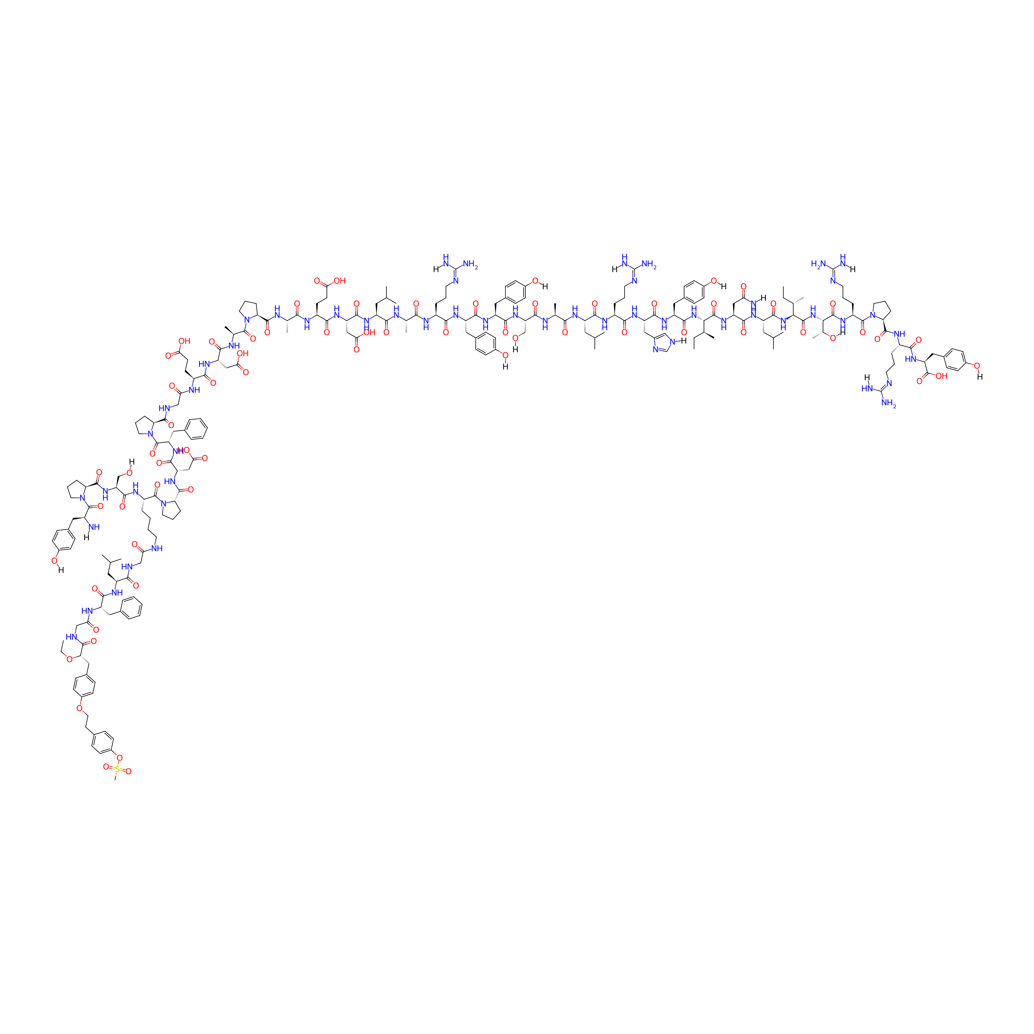
|
|||||
| Peptide Name |
[K4(C-βA-),F7,L17,P34]-hNPY
|
Peptide Info | ||||
| Receptor Name |
Neuropeptide Y receptor type 1 (NPY1R)
|
Receptor Info | ||||
| Drug Name |
Tesaglitazar
|
Drug Info | ||||
| Therapeutic Target |
Peroxisome proliferator-activated receptor alpha (PPARA)
|
Target Info | ||||
| Linker Name |
GFLG
|
Linker Info | ||||
| Peptide Modified Type |
Amino acid modifications
|
|||||
| Modified Segment |
7-Phenylalanine;34-Proline
|
|||||
| Formula |
C234H336N56O66S
|
|||||
| #Ro5 Violations (Lipinski): 5 | Molecular Weight | 5021.655 | ||||
| Lipid-water partition coefficient (xlogp) | -12.2173 | |||||
| Hydrogen Bond Donor Count (hbonddonor) | 60 | |||||
| Hydrogen Bond Acceptor Count (hbondacc) | 66 | |||||
| Rotatable Bond Count (rotbonds) | 160 | |||||
Full List of Activity Data of This Peptide-drug Conjugate
Obtained from the Model Organism Data
| Experiment 1 Reporting the Activity Data of This PDC | [1] | ||||
| Indication | Type 2 diabetes | ||||
| Efficacy Data | Body weigth change |
10%
|
|||
| Administration Time | Every day; over 8 days | ||||
| Administration Dosage | 2.5 µM | ||||
| Evaluation Method | Body weigth change assay | ||||
| MOA of PDC |
In vitro studies revealed that the tesaglitazar-[F7, P34]-NPY conjugate selectively activates PPARγ in NPY7R-expressing cells and enhances adipocyte differentiation and adiponectin expression in adipocyte precursor cells.Additionally, tesa-NPY induces adipocyte differentiation in vivo.
|
||||
| Description |
The mice treated with tesa and tesa-NPY (3) did not change significantly, whereas their littermates treated with [F7, P34]-NPY (2) or vehicle/untreated lost approximately 3% of their body weight (Figure 7A).
|
||||
| In Vivo Model | db/db mice model. | ||||
| Experiment 2 Reporting the Activity Data of This PDC | [1] | ||||
| Indication | Type 2 diabetes | ||||
| Efficacy Data | Cholesterol level |
2.8
|
|||
| Administration Time | Every day; over 8 days | ||||
| Administration Dosage | 2.5 µM | ||||
| MOA of PDC |
In vitro studies revealed that the tesaglitazar-[F7, P34]-NPY conjugate selectively activates PPARγ in NPY1R-expressing cells and enhances adipocyte differentiation and adiponectin expression in adipocyte precursor cells.Additionally, tesa-NPY induces adipocyte differentiation in vivo.
|
||||
| Description |
The influence of tesa, tesa-NPY (3), and [F7, P34]-NPY (2) on the plasma lipids was also analyzed (Figure 9). The vehicle/untreated db/db mice showed elevated levels of triglycerides and free fatty acids (FFA) compared to the lean C57BL/6N mice. Treatment with tesa and tesa-NPY (3) led to a normalization of the triglycerides, FFA, whereas [F7, P34]-NPY (2) and vehicle/untreated had no influence on the lipid metabolism (Figure 9A/B). The cholesterol levels were unchanged by any treatment as these levels were also comparable in the untreated db/db mice compared to the lean mice (Figure 9C).
Click to Show/Hide
|
||||
| In Vivo Model | db/db mice model. | ||||
| Experiment 3 Reporting the Activity Data of This PDC | [1] | ||||
| Indication | Type 2 diabetes | ||||
| Efficacy Data | HbA1C change |
1.05%
|
|||
| Administration Time | Every day; over 8 days | ||||
| Administration Dosage | 2.5 µM | ||||
| MOA of PDC |
In vitro studies revealed that the tesaglitazar-[F7, P34]-NPY conjugate selectively activates PPARγ in NPY1R-expressing cells and enhances adipocyte differentiation and adiponectin expression in adipocyte precursor cells.Additionally, tesa-NPY induces adipocyte differentiation in vivo.
|
||||
| Description |
Whereas in the vehicle/untreated mice, the HbA1C values increased by approximately 2%, a graduated reduced increase was seen for [F7, P34]-NPY (2), peptide conjugate (3), and tesa (Figure 8A). Body temperature, which decreased to 35 C in the vehicle/untreated db/db controls, was normalized to 36 C in all of the treated mice including the mice treated with [F7, P34]-NPY (2) (Figure 8B). Treatment with tesa and tesa-NPY (3) led to normalization of the plasma concentration of ketone bodies and adiponectin, whereas [F7, P34]-NPY (2) and vehicle/untreated showed no effect (Figure 8C/E). Treatment had no major influence on the insulin and Mcp-1 levels (Figure 8D/G). The serum leptin concentration was reduced in the mice treated with tesa, whereas no reduction was detectable in all of the other treated mice (Figure 8F).
Click to Show/Hide
|
||||
| In Vivo Model | db/db mice model. | ||||
| Experiment 4 Reporting the Activity Data of This PDC | [1] | ||||
| Indication | Type 2 diabetes | ||||
| Efficacy Data | Insulin level |
2 ng/mL
|
|||
| Administration Time | Every day; over 8 days | ||||
| Administration Dosage | 2.5 µM | ||||
| MOA of PDC |
In vitro studies revealed that the tesaglitazar-[F7, P34]-NPY conjugate selectively activates PPARγ in NPY1R-expressing cells and enhances adipocyte differentiation and adiponectin expression in adipocyte precursor cells.Additionally, tesa-NPY induces adipocyte differentiation in vivo.
|
||||
| Description |
Whereas in the vehicle/untreated mice, the HbA1C values increased by approximately 2%, a graduated reduced increase was seen for [F7, P34]-NPY (2), peptide conjugate (3), and tesa (Figure 8A). Body temperature, which decreased to 35 C in the vehicle/untreated db/db controls, was normalized to 36 C in all of the treated mice including the mice treated with [F7, P34]-NPY (2) (Figure 8B). Treatment with tesa and tesa-NPY (3) led to normalization of the plasma concentration of ketone bodies and adiponectin, whereas [F7, P34]-NPY (2) and vehicle/untreated showed no effect (Figure 8C/E). Treatment had no major influence on the insulin and Mcp-1 levels (Figure 8D/G). The serum leptin concentration was reduced in the mice treated with tesa, whereas no reduction was detectable in all of the other treated mice (Figure 8F).
Click to Show/Hide
|
||||
| In Vivo Model | db/db mice model. | ||||
| Experiment 5 Reporting the Activity Data of This PDC | [1] | ||||
| Indication | Type 2 diabetes | ||||
| Efficacy Data | Ketone level |
0.5 mM/L
|
|||
| Administration Time | Every day; over 8 days | ||||
| Administration Dosage | 2.5 µM | ||||
| MOA of PDC |
In vitro studies revealed that the tesaglitazar-[F7, P34]-NPY conjugate selectively activates PPARγ in NPY1R-expressing cells and enhances adipocyte differentiation and adiponectin expression in adipocyte precursor cells.Additionally, tesa-NPY induces adipocyte differentiation in vivo.
|
||||
| Description |
Whereas in the vehicle/untreated mice, the HbA1C values increased by approximately 2%, a graduated reduced increase was seen for [F7, P34]-NPY (2), peptide conjugate (3), and tesa (Figure 8A). Body temperature, which decreased to 35 C in the vehicle/untreated db/db controls, was normalized to 36 C in all of the treated mice including the mice treated with [F7, P34]-NPY (2) (Figure 8B). Treatment with tesa and tesa-NPY (3) led to normalization of the plasma concentration of ketone bodies and adiponectin, whereas [F7, P34]-NPY (2) and vehicle/untreated showed no effect (Figure 8C/E). Treatment had no major influence on the insulin and Mcp-1 levels (Figure 8D/G). The serum leptin concentration was reduced in the mice treated with tesa, whereas no reduction was detectable in all of the other treated mice (Figure 8F).
Click to Show/Hide
|
||||
| In Vivo Model | db/db mice model. | ||||
| Experiment 6 Reporting the Activity Data of This PDC | [1] | ||||
| Indication | Type 2 diabetes | ||||
| Efficacy Data | Leptin level |
82 ng/mL
|
|||
| Administration Time | Every day; over 8 days | ||||
| Administration Dosage | 2.5 µM | ||||
| MOA of PDC |
In vitro studies revealed that the tesaglitazar-[F7, P34]-NPY conjugate selectively activates PPARγ in NPY1R-expressing cells and enhances adipocyte differentiation and adiponectin expression in adipocyte precursor cells.Additionally, tesa-NPY induces adipocyte differentiation in vivo.
|
||||
| Description |
Whereas in the vehicle/untreated mice, the HbA1C values increased by approximately 2%, a graduated reduced increase was seen for [F7, P34]-NPY (2), peptide conjugate (3), and tesa (Figure 8A). Body temperature, which decreased to 35 C in the vehicle/untreated db/db controls, was normalized to 36 C in all of the treated mice including the mice treated with [F7, P34]-NPY (2) (Figure 8B). Treatment with tesa and tesa-NPY (3) led to normalization of the plasma concentration of ketone bodies and adiponectin, whereas [F7, P34]-NPY (2) and vehicle/untreated showed no effect (Figure 8C/E). Treatment had no major influence on the insulin and Mcp-1 levels (Figure 8D/G). The serum leptin concentration was reduced in the mice treated with tesa, whereas no reduction was detectable in all of the other treated mice (Figure 8F).
Click to Show/Hide
|
||||
| In Vivo Model | db/db mice model. | ||||
| Experiment 7 Reporting the Activity Data of This PDC | [1] | ||||
| Indication | Type 2 diabetes | ||||
| Efficacy Data | Mcp-1 level |
305 pg/mL
|
|||
| Administration Time | Every day; over 8 days | ||||
| Administration Dosage | 2.5 µM | ||||
| MOA of PDC |
In vitro studies revealed that the tesaglitazar-[F7, P34]-NPY conjugate selectively activates PPARγ in NPY1R-expressing cells and enhances adipocyte differentiation and adiponectin expression in adipocyte precursor cells.Additionally, tesa-NPY induces adipocyte differentiation in vivo.
|
||||
| Description |
Whereas in the vehicle/untreated mice, the HbA1C values increased by approximately 2%, a graduated reduced increase was seen for [F7, P34]-NPY (2), peptide conjugate (3), and tesa (Figure 8A). Body temperature, which decreased to 35 C in the vehicle/untreated db/db controls, was normalized to 36 C in all of the treated mice including the mice treated with [F7, P34]-NPY (2) (Figure 8B). Treatment with tesa and tesa-NPY (3) led to normalization of the plasma concentration of ketone bodies and adiponectin, whereas [F7, P34]-NPY (2) and vehicle/untreated showed no effect (Figure 8C/E). Treatment had no major influence on the insulin and Mcp-1 levels (Figure 8D/G). The serum leptin concentration was reduced in the mice treated with tesa, whereas no reduction was detectable in all of the other treated mice (Figure 8F).
Click to Show/Hide
|
||||
| In Vivo Model | db/db mice model. | ||||
| Experiment 8 Reporting the Activity Data of This PDC | [1] | ||||
| Indication | Type 2 diabetes | ||||
| Efficacy Data | Triglycerides level |
0.65
|
|||
| Administration Time | Every day; over 8 days | ||||
| Administration Dosage | 2.5 µM | ||||
| MOA of PDC |
In vitro studies revealed that the tesaglitazar-[F7, P34]-NPY conjugate selectively activates PPARγ in NPY1R-expressing cells and enhances adipocyte differentiation and adiponectin expression in adipocyte precursor cells.Additionally, tesa-NPY induces adipocyte differentiation in vivo.
|
||||
| Description |
The influence of tesa, tesa-NPY (3), and [F7, P34]-NPY (2) on the plasma lipids was also analyzed (Figure 9). The vehicle/untreated db/db mice showed elevated levels of triglycerides and free fatty acids (FFA) compared to the lean C57BL/6N mice. Treatment with tesa and tesa-NPY (3) led to a normalization of the triglycerides, FFA, whereas [F7, P34]-NPY (2) and vehicle/untreated had no influence on the lipid metabolism (Figure 9A/B). The cholesterol levels were unchanged by any treatment as these levels were also comparable in the untreated db/db mice compared to the lean mice (Figure 9C).
Click to Show/Hide
|
||||
| In Vivo Model | db/db mice model. | ||||
| Experiment 9 Reporting the Activity Data of This PDC | [1] | ||||
| Indication | Type 2 diabetes | ||||
| Efficacy Data | Triglycerides level |
0.9
|
|||
| Administration Time | Every day; over 8 days | ||||
| Administration Dosage | 2.5 µM | ||||
| MOA of PDC |
In vitro studies revealed that the tesaglitazar-[F7, P34]-NPY conjugate selectively activates PPARγ in NPY1R-expressing cells and enhances adipocyte differentiation and adiponectin expression in adipocyte precursor cells.Additionally, tesa-NPY induces adipocyte differentiation in vivo.
|
||||
| Description |
The influence of tesa, tesa-NPY (3), and [F7, P34]-NPY (2) on the plasma lipids was also analyzed (Figure 9). The vehicle/untreated db/db mice showed elevated levels of triglycerides and free fatty acids (FFA) compared to the lean C57BL/6N mice. Treatment with tesa and tesa-NPY (3) led to a normalization of the triglycerides, FFA, whereas [F7, P34]-NPY (2) and vehicle/untreated had no influence on the lipid metabolism (Figure 9A/B). The cholesterol levels were unchanged by any treatment as these levels were also comparable in the untreated db/db mice compared to the lean mice (Figure 9C).
Click to Show/Hide
|
||||
| In Vivo Model | db/db mice model. | ||||
References